This paper proposes a practical method for optimizing CBCT dose using simple measurement equipment, demonstrating that significant dose reductions (up to 80%) can be achieved while maintaining acceptable image quality, facilitating straightforward implementation in radiation oncology departments.
Objective: To propose a CBCT dose optimization method using readily available measurement equipment in radiation oncology departments. 
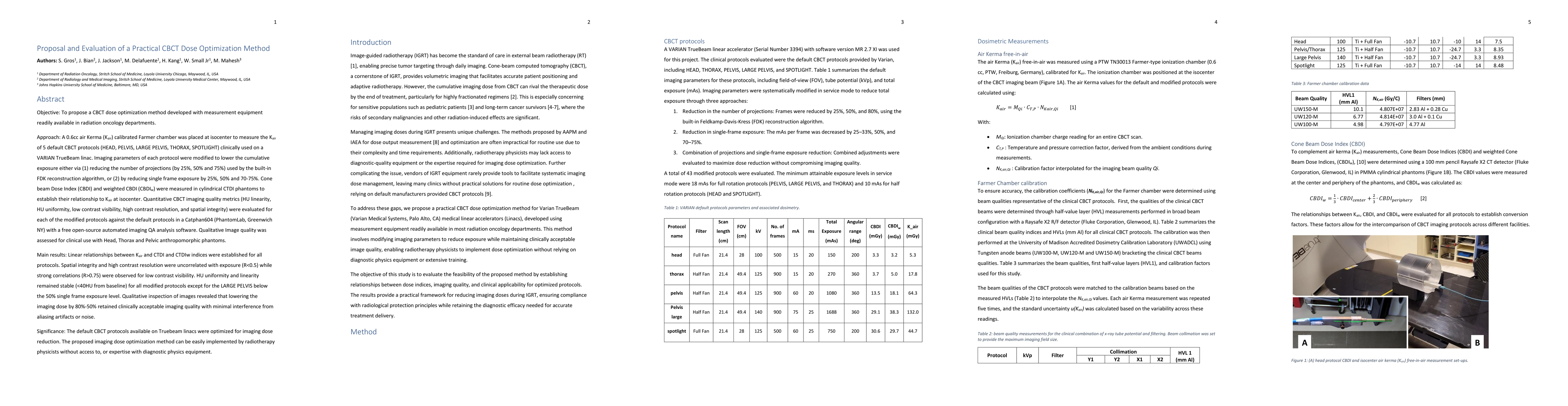
Approach: A 0.6cc air kerma (Kair) calibrated Farmer chamber measured Kair at isocenter for five default CBCT protocols (HEAD, PELVIS, LARGE PELVIS, THORAX, SPOTLIGHT) on a Varian TrueBeam linac. Imaging parameters were modified to reduce cumulative exposure by reducing projections (by 25%, 50%, 75%) in FDK reconstruction, or lowering single frame exposure by 25%, 50%, 70 to 75%. Cone Beam Dose Index (CBDI) and weighted CBDI (CBDI_w) were measured in CTDI phantoms to correlate with Kair. Image quality was assessed quantitatively (HU linearity, uniformity, low-contrast visibility, high-contrast resolution, spatial integrity) using Catphan604, and qualitatively via anthropomorphic phantoms. Results: A linear relationship between Kair, CTDI, and CBDI_w was established. High-contrast resolution and spatial integrity were unaffected by dose reduction (R < 0.5), while low-contrast visibility strongly correlated with exposure (R > 0.75). HU uniformity and linearity remained stable (<40 HU deviation), except for LARGE PELVIS at <50% single-frame exposure. 80 to 50% dose reduction retained clinically acceptable image quality with minimal artifacts or noise. Significance: CBCT protocols on TrueBeam linacs were optimized for dose reduction. The method enables easy implementation by radiotherapy physicists without diagnostic physics equipment or expertise.
Generated Jun 12, 2025
The research proposes a CBCT dose optimization method using a Farmer chamber to measure air kerma (Kair) for five default CBCT protocols on a Varian TrueBeam linac. Imaging parameters are modified to reduce cumulative exposure by decreasing projections or lowering single-frame exposure. Image quality is assessed quantitatively and qualitatively.
This method enables easy implementation by radiotherapy physicists without diagnostic physics equipment or expertise, significantly reducing CBCT doses while maintaining clinically acceptable image quality, especially beneficial for pediatric patients.
The study establishes a practical approach to CBCT dose optimization using readily available measurement equipment, demonstrating substantial dose reductions without significant degradation in quantitative imaging quality.
This work bridges the gap between previous studies by demonstrating clinically viable dose reductions across multiple acquisition modes and developing institution-specific protocols, including a successful pediatric implementation for neuroblastoma IGRT.
No key terms available for this paper.
Current paper (gray), citations (green), references (blue)
Farhang Bayat, Brian Miller, Yeonok Park et al.
Ying Yuan, Heng Zhou, Suyu Liu
Louise Friot-Giroux, Françoise Peyrin, Voichita Maxim
No citations found for this paper.
Comments (0)