Prostate cancer inference via weakly-supervised learning using a large collection of negative MRI
Publication
Metrics
Paper Preview
Abstract
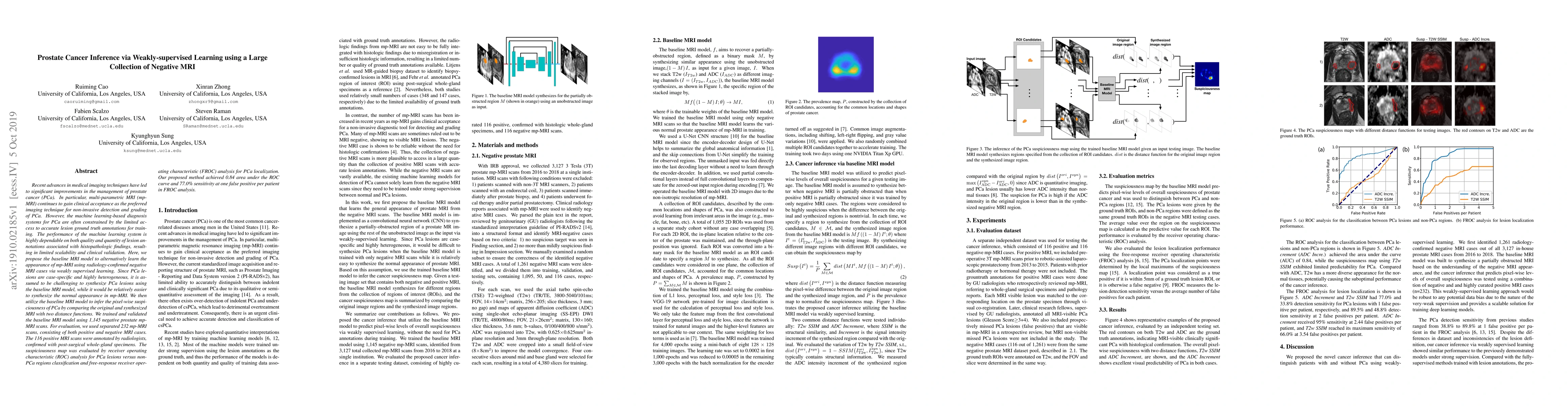
Recent advances in medical imaging techniques have led to significant improvements in the management of prostate cancer (PCa). In particular, multi-parametric MRI (mp-MRI) continues to gain clinical acceptance as the preferred imaging technique for non-invasive detection and grading of PCa. However, the machine learning-based diagnosis systems for PCa are often constrained by the limited access to accurate lesion ground truth annotations for training. The performance of the machine learning system is highly dependable on both quality and quantity of lesion annotations associated with histopathologic findings, resulting in limited scalability and clinical validation. Here, we propose the baseline MRI model to alternatively learn the appearance of mp-MRI using radiology-confirmed negative MRI cases via weakly supervised learning. Since PCa lesions are case-specific and highly heterogeneous, it is assumed to be challenging to synthesize PCa lesions using the baseline MRI model, while it would be relatively easier to synthesize the normal appearance in mp-MRI. We then utilize the baseline MRI model to infer the pixel-wise suspiciousness of PCa by comparing the original and synthesized MRI with two distance functions. We trained and validated the baseline MRI model using 1,145 negative prostate mp-MRI scans. For evaluation, we used separated 232 mp-MRI scans, consisting of both positive and negative MRI cases. The 116 positive MRI scans were annotated by radiologists, confirmed with post-surgical whole-gland specimens. The suspiciousness map was evaluated by receiver operating characteristic (ROC) analysis for PCa lesions versus non-PCa regions classification and free-response receiver operating characteristic (FROC) analysis for PCa localization. Our proposed method achieved 0.84 area under the ROC curve and 77.0% sensitivity at one false positive per patient in FROC analysis.
AI Key Findings
Get AI-generated insights about this paper's methodology, results, significance, and more — seven facets brought into focus.
Impact
Paper Details
Authors
PDF Preview
Key Terms
Citation Network
Current paper (gray), citations (green), references (blue)
Display is limited for performance on very large graphs.
Discussion 0