Protein-protein modelling using cryo-EM restraints
Publication
Metrics
AI Quick Summary
A new protocol uses cryo-EM restraints to improve protein-protein modelling, accounting for molecular recognition, energy, and flexibility in addition to traditional rigid fitting methods.
Paper Preview
Abstract
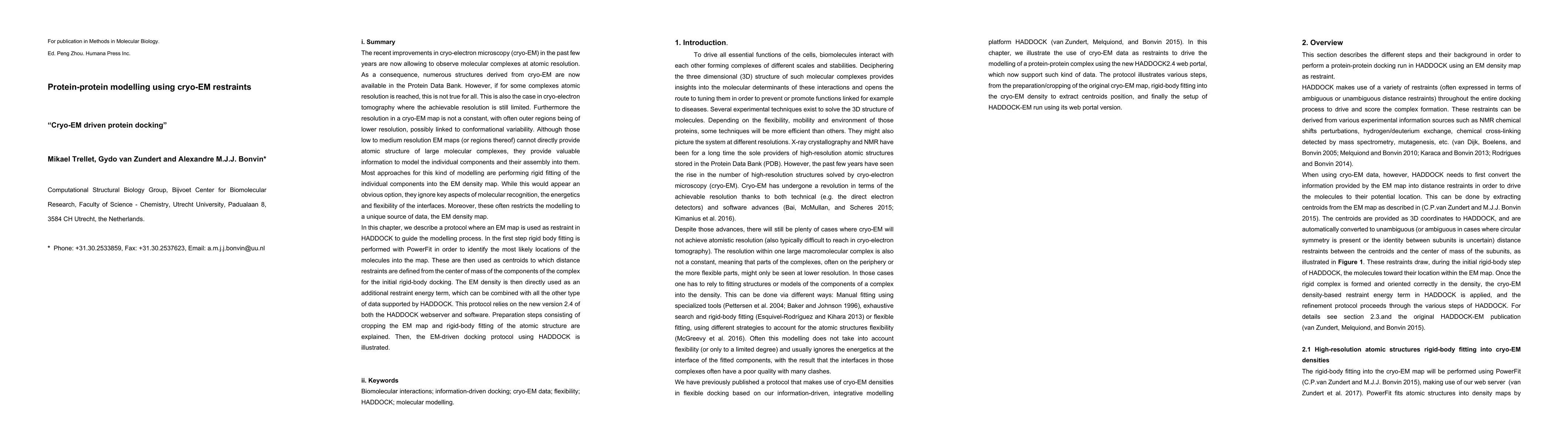
The recent improvements in cryo-electron microscopy (cryo-EM) in the past few years are now allowing to observe molecular complexes at atomic resolution. As a consequence, numerous structures derived from cryo-EM are now available in the Protein Data Bank. However, if for some complexes atomic resolution is reached, this is not true for all. This is also the case in cryo-electron tomography where the achievable resolution is still limited. Furthermore the resolution in a cryo-EM map is not a constant, with often outer regions being of lower resolution, possibly linked to conformational variability. Although those low to medium resolution EM maps (or regions thereof) cannot directly provide atomic structure of large molecular complexes, they provide valuable information to model the individual components and their assembly into them. Most approaches for this kind of modelling are performing rigid fitting of the individual components into the EM density map. While this would appear an obvious option, they ignore key aspects of molecular recognition, the energetics and flexibility of the interfaces. Moreover, these often restricts the modelling to a unique source of data, the EM density map. In this chapter, we describe a protocol where an EM map is used as restraint in HADDOCK to guide the modelling process.
AI Key Findings
Get AI-generated insights about this paper's methodology, results, significance, and more — seven facets brought into focus.
Impact
Paper Details
Authors
PDF Preview
Key Terms
Citation Network
Current paper (gray), citations (green), references (blue)
Display is limited for performance on very large graphs.
Discussion 0