Publication
Metrics
AI Quick Summary
This paper demonstrates that bright-field differential dynamic microscopy can accurately determine the size and diffusion coefficient of small macromolecules like proteins, even at low concentrations, rivaling the precision of commercial dynamic light scattering systems. The method offers a rapid, label-free alternative for protein sizing using an optical microscope.
Paper Preview
Abstract
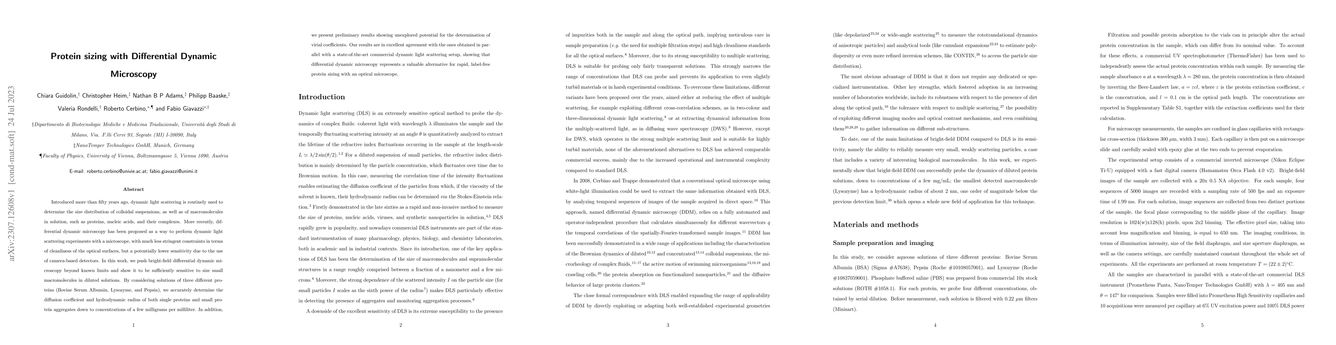
Introduced more than fifty years ago, dynamic light scattering is routinely used to determine the size distribution of colloidal suspensions, as well as of macromolecules in solution, such as proteins, nucleic acids, and their complexes. More recently, differential dynamic microscopy has been proposed as a way to perform dynamic light scattering experiments with a microscope, with much less stringent constraints in terms of cleanliness of the optical surfaces, but a potentially lower sensitivity due to the use of camera-based detectors. In this work, we push bright-field differential dynamic microscopy beyond known limits and show it to be sufficiently sensitive to size small macromolecules in diluted solutions. By considering solutions of three different proteins (Bovine Serum Albumin, Lysozyme, and Pepsin), we accurately determine the diffusion coefficient and hydrodynamic radius of both single proteins and small protein aggregates down to concentrations of a few milligrams per milliliter. In addition, we present preliminary results showing unexplored potential for the determination of virial coefficients. Our results are in excellent agreement with the ones obtained in parallel with a state-of-the-art commercial dynamic light scattering setup, showing that differential dynamic microscopy represents a valuable alternative for rapid, label-free protein sizing with an optical microscope.
AI Key Findings
Get AI-generated insights about this paper's methodology, results, significance, and more — seven facets brought into focus.
Impact
Paper Details
Authors
PDF Preview
Key Terms
Citation Network
Current paper (gray), citations (green), references (blue)
Display is limited for performance on very large graphs.
Discussion 0