Publication
Metrics
AI Quick Summary
A new proton radiography scanner prototype uses position-sensitive silicon detectors and high-resolution scintillators to produce images of materials with proton beams. It successfully resolves structures up to 2 mm and distinguishes thicknesses of up to 10 mm.
Paper Preview
Abstract
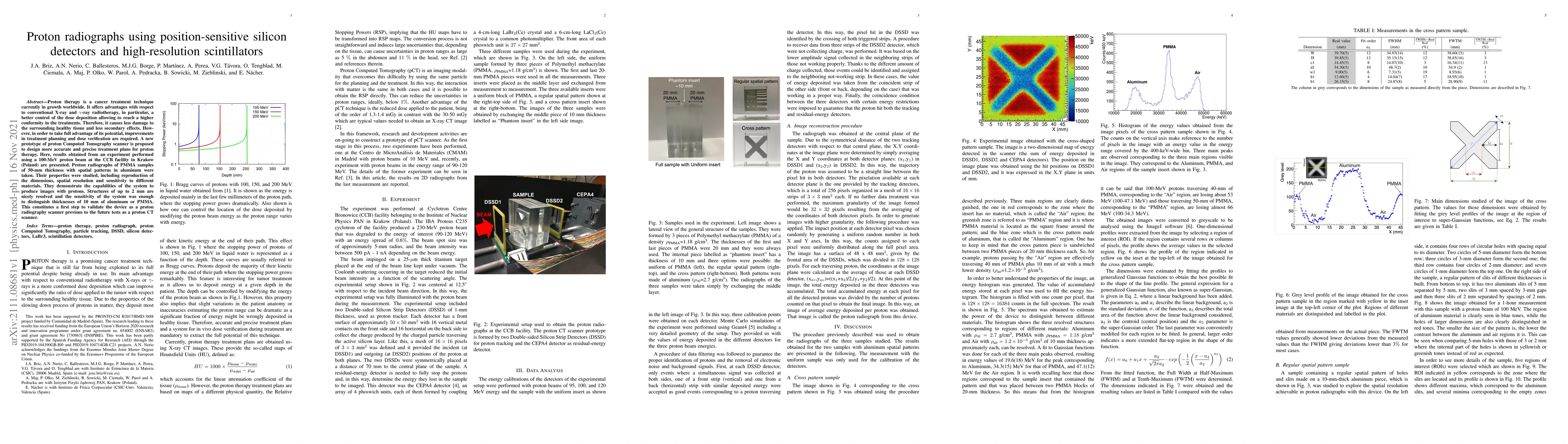
Proton therapy is a cancer treatment technique currently in growth worldwide. It offers advantages with respect to conventional X-ray and $\gamma$-ray radiotherapy, in particular, a better control of the dose deposition allowing to reach a higher conformity in the treatments. Therefore, it causes less damage to the surrounding healthy tissue and less secondary effects. However, in order to take full advantage of its potential, improvements in treatment planning and dose verification are required. A new prototype of proton Computed Tomography scanner is proposed to design more accurate and precise treatment plans for proton therapy. Here, results obtained from an experiment performed using a 100-MeV proton beam at the CCB facility in Krakow (Poland) are presented. Proton radiographs of PMMA samples of 50-mm thickness with spatial patterns in aluminum were taken. Their properties were studied, including reproduction of the dimensions, spatial resolution and sensitivity to different materials. They demonstrate the capabilities of the system to produce images with protons. Structures of up to 2 mm are nicely resolved and the sensitivity of the system was enough to distinguish thicknesses of 10 mm of aluminum or PMMA. This constitutes a first step to validate the device as a proton radiography scanner previous to the future tests as a proton CT scanner.
AI Key Findings
Get AI-generated insights about this paper's methodology, results, significance, and more — seven facets brought into focus.
Impact
Paper Details
Authors
PDF Preview
Key Terms
Citation Network
Current paper (gray), citations (green), references (blue)
Display is limited for performance on very large graphs.
Discussion 0