PseudoCell: Hard Negative Mining as Pseudo Labeling for Deep Learning-Based Centroblast Cell Detection
Publication
Metrics
AI Quick Summary
PseudoCell automates centroblast cell detection in whole-slide images using deep learning, reducing pathologists' workload by eliminating 58.18-99.35% of non-centroblast areas through pseudo-negative mining and morphological features. This framework requires initial pathologist labels but does not need continuous refinement for performance improvement.
Paper Preview
Abstract
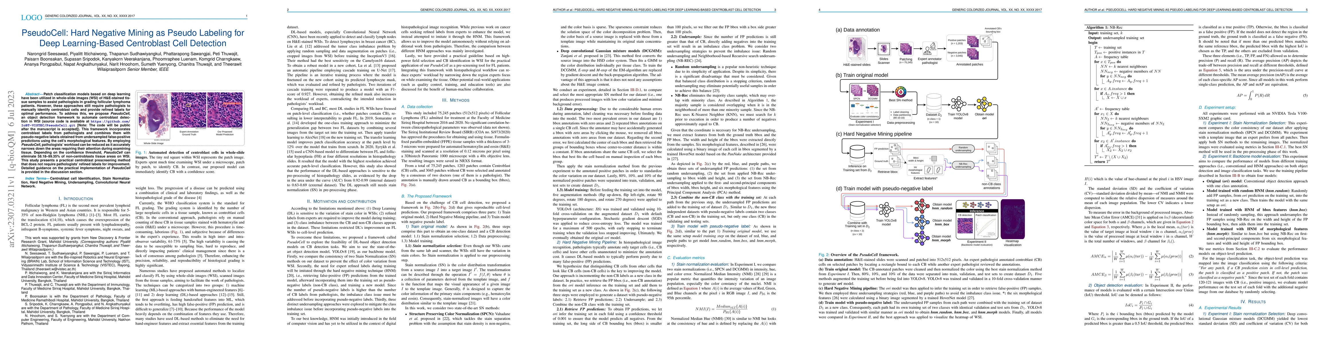
Patch classification models based on deep learning have been utilized in whole-slide images (WSI) of H&E-stained tissue samples to assist pathologists in grading follicular lymphoma patients. However, these approaches still require pathologists to manually identify centroblast cells and provide refined labels for optimal performance. To address this, we propose PseudoCell, an object detection framework to automate centroblast detection in WSI (source code is available at https://github.com/IoBT-VISTEC/PseudoCell.git). This framework incorporates centroblast labels from pathologists and combines them with pseudo-negative labels obtained from undersampled false-positive predictions using the cell's morphological features. By employing PseudoCell, pathologists' workload can be reduced as it accurately narrows down the areas requiring their attention during examining tissue. Depending on the confidence threshold, PseudoCell can eliminate 58.18-99.35% of non-centroblasts tissue areas on WSI. This study presents a practical centroblast prescreening method that does not require pathologists' refined labels for improvement. Detailed guidance on the practical implementation of PseudoCell is provided in the discussion section.
AI Key Findings
Get AI-generated insights about this paper's methodology, results, significance, and more — seven facets brought into focus.
Impact
Paper Details
Authors
PDF Preview
Key Terms
Citation Network
Current paper (gray), citations (green), references (blue)
Display is limited for performance on very large graphs.
Discussion 0