Pycro-Manager is an open-source software that integrates microscopy hardware control and image processing, bridging the gap between {\mu}Manager's C++/Java libraries and Python's data science tools for more intuitive and efficient experiments. It facilitates rapid development and leverages existing {\mu}Manager capabilities through Python.
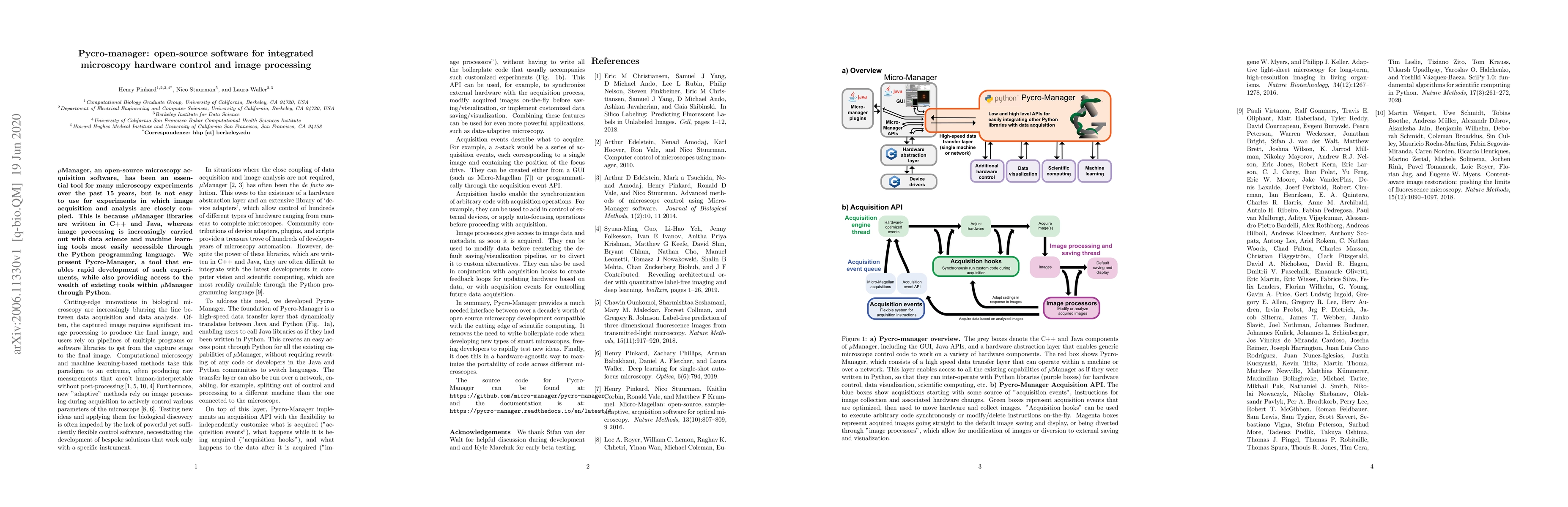
{\mu}Manager, an open-source microscopy acquisition software, has been an essential tool for many microscopy experiments over the past 15 years, but is not easy to use for experiments in which image acquisition and analysis are closely coupled. This is because {\mu}Manager libraries are written in C++ and Java, whereas image processing is increasingly carried out with data science and machine learning tools most easily accessible through the Python programming language. We present Pycro-Manager, a tool that enables rapid development of such experiments, while also providing access to the wealth of existing tools within {\mu}Manager through Python.
Get AI-generated insights about this paper's methodology, results, and significance.
Current paper (gray), citations (green), references (blue)
Marco Guarnieri, Gideon Mohr, Jan Reineke
Title | Authors | Year | Actions |
---|---|---|---|
A novel expression system for imaging single-molecule fluorescence in Haloferax volcanii WR806 enables visualization of Cas1 involvement in UV-light associated DNA repair
|
Paula R Schrage, U. Afonina, Julia Wörtz, A. Marchfelder, Koen J. A. Martens, J. Sáenz, Ulrike Endesfelder
|
2025
|
View Paper |
DMC-BrainMap - an open-source, end-to-end tool for multi-feature brain mapping across species
|
Felix Jung, Xiao Cao, Loran Heymans, M. Carlén
|
2025
|
View Paper |
Teach your microscope how to print: Low-cost and rapid-iteration microfabrication for biology
|
Lucien Hinderling, Remo Hadorn, Moritz Kwasny, Joël Frei, Benjamin Grädel, Sacha Psalmon, Yannick Blum, Rémi Berthoz, Alex Landolt, Benjamin D. Towbin, Daniel Riveline, Olivier Pertz
|
2025
|
View Paper |
Self-Driving Microscopes: AI Meets Super-Resolution Microscopy.
|
Edward N. Ward, Anna Scheeder, Max Barysevich, C. Kaminski
|
2025
|
View Paper |
Single-shot label-free nanoscopy for quantitative organelle visualization on standard commercial microscopes
|
Yugo Inutsuka, Koki Yamamoto, Masafumi Kuroda, Yasushi Okada
|
2025
|
View Paper |
Replicable and extensible spatial data acquisition
|
Matthew Haffner
|
2024
|
View Paper |
Machine learning in microscopy - insights, opportunities and challenges.
|
Inês M. Cunha, Emma Latron, Sebastian Bauer, Daniel Sage, Juliette Griffié
|
2024
|
View Paper |
The eduWOSM: a benchtop advanced microscope for education and research
|
N.J. Carter, D.S. Martin, J. E. Molloy, R. A. Cross
|
2024
|
View Paper |
The Roboscope: Smart and Fast Microscopy for Generic Event-Driven Acquisition
|
Julia Bonnet, Youssef El-Habouz, Célia Martin, Maelle Guillout, Louis Ruel, Baptiste Giroux, Claire Déméautis, Benjamin Mercat, Otmane Bouchareb, J. Pécréaux, Marc Tramier
|
2024
|
View Paper |
ClearScope: a fully integrated light sheet theta microscope for sub-cellular resolution imaging without lateral size constraints
|
Matthew G. Fay, Peter J. Lang, David S. Denu, Nathan J. O’Connor, Benjamin Haydock, Jeffrey Blaisdell, Nicolas Roussel, Alissa Wilson, S. M. Aronson, Paul J. Angstman, Cheng Gong, Tanvi Butola, O. Devinsky, Jayeeta Basu, Raju Tomer, Jacob R. Glaser
|
2024
|
View Paper |
Mantis: High-throughput 4D imaging and analysis of the molecular and physical architecture of cells
|
Ivan E. Ivanov, Eduardo Hirata-Miyasaki, Talon Chandler, Rasmi Cheloor-Kovilakam, Ziwen Liu, Soorya Pradeep, Chad Liu, Madhura Bhave, Sudip Khadka, Carolina Arias, Manuel D. Leonetti, Bo Huang, Shalin B. Mehta
|
2024
|
View Paper |
A DIY guide for image-based spatial transcriptomic: TLS as a case example
|
Thomas Defard, Auxence Desrentes, C. Fouillade, Florian Mueller
|
2024
|
View Paper |
Scanless two-photon voltage imaging
|
Ruth R. Sims, Imane Bendifallah, Christiane Grimm, A. Lafirdeen, Soledad Domínguez, Chung Yuen Chan, Xiaoyu Lu, Benoît C Forget, François St-Pierre, E. Papagiakoumou, Valentina Emiliani
|
2024
|
View Paper |
Innovating in a bioimaging core through instrument development
|
Sebastian Munck, Christof De Bo, Christopher J Cawthorne, Julien Colombelli
|
2024
|
View Paper |
ZOLA 3D, single molecule microscope technological transfer from research lab to ISO 9001 core facility
|
Yumi Gosselin, M. Lelek, Audrey Salles, Christophe Zimmer, Spencer L. Shorte, Elric Esposito
|
2024
|
View Paper |
The Berkeley Single Cell Computational Microscopy (BSCCM) Dataset
|
H. Pinkard, Cherry Liu, Fanice Nyatigo, Daniel A. Fletcher, Laura Waller
|
2024
|
View Paper |
Multi-domain automated patterning of DNA-functionalized hydrogels
|
Moshe Rubanov, Joshua Cole, H. Lee, Leandro G. Soto Cordova, Zachary Chen, Elia Gonzalez, Rebecca Schulman
|
2024
|
View Paper |
AEcroscopy: A Software-Hardware Framework Empowering Microscopy Toward Automated and Autonomous Experimentation.
|
Yongtao Liu, Kevin M. Roccapriore, M. Checa, Sai Mani Prudhvi Valleti, Jan-Chi Yang, Stephen Jesse, R. Vasudevan
|
2023
|
View Paper |
Aberration correction for deformable mirror based remote focusing enables high-accuracy whole-cell super-resolution imaging
|
Wei Shi, Yingchuan He, Jianlin Wang, Lulu Zhou, Jianwei Chen, Liwei Zhou, Zeyu Xi, Zhen Wang, Ke Fang, Yiming Li
|
2023
|
View Paper |
Live-cell imaging in the deep learning era.
|
Joanna W. Pylvänäinen, Estibaliz Gómez-de-Mariscal, Ricardo Henriques, Guillaume Jacquemet
|
2023
|
View Paper |
Nucleolar dynamics are determined by the ordered assembly of the ribosome
|
Jessica Sheu-Gruttadauria, Xiaowei Yan, N. Stuurman, Ronald D. Vale, S. Floor
|
2023
|
View Paper |
Local negative feedback of Rac activity at the leading edge underlies a pilot pseudopod-like program for amoeboid cell guidance
|
J. Town, O. Weiner
|
2023
|
View Paper |
Temporal analysis of relative distances (TARDIS) is a robust, parameter-free alternative to single-particle tracking
|
Koen J. A. Martens, Bartosz Turkowyd, J. Hohlbein, Ulrike Endesfelder
|
2023
|
View Paper |
Light-sheets and smart microscopy, an exciting future is dawning
|
S. Daetwyler, R. Fiolka
|
2023
|
View Paper |
Pupil engineering for extended depth-of-field imaging in a fluorescence miniscope
|
Joseph Greene, Yujia Xue, Jeffrey Alido, Alex Matlock, Guo-ying Hu, K. Kılıç, I. Davison, L. Tian
|
2023
|
View Paper |
Probe microscopy is all you need
|
S. Kalinin, R. Vasudevan, Yongtao Liu, Ayana Ghosh, Kevin M. Roccapriore, M. Ziatdinov
|
2023
|
View Paper |
Data-driven microscopy allows for automated context-specific acquisition of high-fidelity image data.
|
Oscar André, Johannes Kumra Ahnlide, Nils Norlin, V. Swaminathan, P. Nordenfelt
|
2023
|
View Paper |
FilamentSensor 2.0: An open-source modular toolbox for 2D/3D cytoskeletal filament tracking
|
L. Hauke, Andreas Primeßnig, B. Eltzner, Jennifer Radwitz, S. Huckemann, F. Rehfeldt
|
2023
|
View Paper |
An open-source microscopy framework for simultaneous control of image acquisition, reconstruction, and analysis
|
Xavier Casas Moreno, Mariline Mendes Silva, Johannes Roos, Francesca Pennacchietti, Nils Norlin, Ilaria Testa
|
2023
|
View Paper |
Hardware-software co-design of an open-source automatic multimodal whole slide histopathology imaging system
|
Bin Li, Mitchell S. Nelson, J. Chacko, Nathan Cudworth, K. Eliceiri
|
2023
|
View Paper |
Scanless two-photon voltage imaging
|
Ruth R. Sims, Imane Bendifallah, C. Grimm, A. Lafirdeen, Xiaoyu Lu, François St-Pierre, E. Papagiakoumou, V. Emiliani
|
2023
|
View Paper |
Rac negative feedback links local PIP3 rate-of-change to dynamic control of neutrophil guidance
|
J. Town, O. Weiner
|
2023
|
View Paper |
Fabrication and validation of an LED array microscope for multimodal, quantitative imaging
|
Tarek E. Moustafa, Edward R. Polanco, R. Belote, Robert Judson-Torres, T. Zangle
|
2023
|
View Paper |
Volumetric imaging of fast cellular dynamics with deep learning enhanced bioluminescence microscopy
|
L. Morales-Curiel, A. Gonzalez, Gustavo Castro-Olvera, Li-Chun Lin, Malak El-Quessny, Montserrat Porta-de-la-Riva, Jacqueline Severino, L. B. Morera, Valeria Venturini, V. Ruprecht, D. Ramallo, P. Loza-Álvarez, Michael Krieg
|
2022
|
View Paper |
Automation of yeast spot assays using an affordable liquid handling robot
|
Shodai Taguchi, Yasuyuki Suda, K. Irie, Haruka Ozaki
|
2022
|
View Paper |
Tracking Mechanical Stress and Cell Migration with Inexpensive Polymer Thin‐Film Sensors
|
Tanner J. Finney, Skye L. Frank, Michael R. Bull, R. Guy, T. Kuhl
|
2022
|
View Paper |
Multiphoton intravital microscopy of rodents
|
C. Scheele, D. Herrmann, Erika Yamashita, Cristina Lo Celso, C. Jenne, M. Oktay, D. Entenberg, P. Friedl, R. Weigert, F. Meijboom, M. Ishii, P. Timpson, J. van Rheenen
|
2022
|
View Paper |
Differentiation of pancreatic ductal adenocarcinoma and chronic pancreatitis using graph neural networks on histopathology and collagen fiber features
|
Bin Li, Michael S. Nelson, Omid Savari, A. Loeffler, K. Eliceiri
|
2022
|
View Paper |
Open-source software package for on-the-fly deskewing and live viewing of volumetric lightsheet microscopy data
|
Jacob R. Lamb, Edward N. Ward, C. Kaminski
|
2022
|
View Paper |
Deep model predictive control of gene expression in thousands of single cells
|
Jean-Baptiste Lugagne, Caroline Blassick, M. Dunlop
|
2022
|
View Paper |
Microscopy is All You Need
|
S. Kalinin, R. Vasudevan, Yongtao Liu, Ayana Ghosh, Kevin M. Roccapriore, M. Ziatdinov
|
2022
|
View Paper |
The miEye: Bench-top super-resolution microscope with cost-effective equipment
|
Mohammad Nour Alsamsam, Aurimas Kopūstas, Meda Jurevičiūtė, Marijonas Tutkus
|
2022
|
View Paper |
Microscopes are coming for your job
|
H. Pinkard, L. Waller
|
2022
|
View Paper |
EDoF-Miniscope: pupil engineering for extended depth-of-field imaging in a fluorescence miniscope
|
Joseph Greene, Yujia Xue, Jeffrey Alido, Alex Matlock, Guo-ying Hu, K. Kılıç, Ian Davison, Lei Tian
|
2022
|
View Paper |
Ring Deconvolution Microscopy: An Exact Solution for Spatially-Varying Aberration Correction
|
Amit Kohli, Anastasios Angelopoulos, David McAllister, Esther Whang, Sixian You, Kyrollos Yanny, Federico M. Gasparoli, L. Waller
|
2022
|
View Paper |
MAI-SIM: interferometric multicolor structured illumination microscopy for everybody
|
Edward N. Ward, Lisa Hecker, Charles N Christensen, Jacob R. Lamb, M. Lu, Luca Mascheroni, C.W. Chung, Anna Wang, C. Rowlands, G. K. Schierle, C. Kaminski
|
2022
|
View Paper |
Volumetric bioluminescence imaging of cellular dynamics with deep learning based light-field reconstruction
|
L. Morales-Curiel, Gustavo Castro-Olvera, A. Gonzalez, Lynn Lin, Malak El-Quessny, Montserrat Porta-de-la-Riva, Jacqueline Severino, Laura Battle, D. Ramallo, V. Ruprecht, P. Loza-Álvarez, Michael Krieg
|
2022
|
View Paper |
A Tailored Approach To Study Legionella Infection Using Lattice Light Sheet Microscope (LLSM)
|
Xiyu Yi, Haichao Miao, Jacky Kai-Yin Lo, Maher M. Elsheikh, T. Lee, Chenfanfu Jiang, Yuliang Zhang, Brent W. Segelke, K. W. Overton, P. Bremer, Ted A. Laurence
|
2022
|
View Paper |
Enabling reactive microscopy with MicroMator
|
Z. Fox, Steven Fletcher, Achille Fraisse, Chetan Aditya, Sebastián Sosa-Carrillo, Julienne Petit, Sébastien Gilles, F. Bertaux, J. Ruess, Grégory Batt
|
2022
|
View Paper |
Live cell microscopy: From image to insight
|
Andreas P. Cuny, Fabian P. Schlottmann, Jennifer C. Ewald, Serge Pelet, K. Schmoller
|
2022
|
View Paper |
A Hitchhiker's guide through the bio‐image analysis software universe
|
R. Haase, E. Fazeli, D. Legland, M. Doube, Siân Culley, I. Belevich, E. Jokitalo, Martin Schorb, Anna H. Klemm, C. Tischer
|
2022
|
View Paper |
DaXi—high-resolution, large imaging volume and multi-view single-objective light-sheet microscopy
|
Bin Yang, Merlin Lange, Alfred Millett-Sikking, Xiangye Zhao, Jordão Bragantini, Shruthi VijayKumar, M. Kamb, Rafael Gómez-Sjöberg, A. Solak, Wanpeng Wang, Hirofumi Kobayashi, Matthew N. McCarroll, L. Whitehead, R. Fiolka, T. Kornberg, Andrew G. York, Loic A. Royer
|
2022
|
View Paper |
Polarization Differential Interference Contrast (PDIC) microscopy for quantitative phase and fluorescence imaging of mouse brain sections
|
Mark Strassberg, Y. Shevtsova, Kai Wagoner-oshima, Chelsea Yu, Jijuan Xu, Christine Ramiro, Jia Liu, Min Xu
|
2022
|
View Paper |
Roadmap on Digital Holography-Based Quantitative Phase Imaging
|
Vinoth Balasubramani, M. Kujawińska, C. Allier, V. Anand, Chau-Jern Cheng, C. Depeursinge, Nathaniel Hai, S. Juodkazis, J. Kalkman, A. Kuś, Moosung Lee, P. Magistretti, P. Marquet, S. Ng, Joseph Rosen, Y. Park, M. Ziemczonok
|
2021
|
View Paper |
Light sheet fluorescence microscopy
|
Ernst H. K. Stelzer, Frederic Strobl, Bo-Jui Chang, Friedrich Preusser, S. Preibisch, Katie McDole, R. Fiolka
|
2021
|
View Paper |
Isotropic imaging across spatial scales with axially swept light-sheet microscopy
|
K. Dean, Tonmoy Chakraborty, S. Daetwyler, Jinlong Lin, Gerard Garrelts, Ons M’Saad, Hannahmariam T. Mekbib, F. Voigt, Martina Schaettin, E. Stoeckli, F. Helmchen, J. Bewersdorf, R. Fiolka
|
2021
|
View Paper |
Open microscopy in the life sciences: quo vadis?
|
J. Hohlbein, Benedict Diederich, Barbora Marsíková, E. Reynaud, S. Holden, W. Jahr, R. Haase, Kirti Prakash
|
2021
|
View Paper |
Characterizing and Tuning the Properties of Polydiacetylene Films for Sensing Applications.
|
Tanner J. Finney, S. Parikh, A. Berman, D. Sasaki, T. Kuhl
|
2021
|
View Paper |
Tissue Engineering with Mechanically Induced Solid‐Fluid Transitions
|
Erik Mailand, Ece Özelçi, Jaemin Kim, Matthias Rüegg, Odysseas Chaliotis, Jon Märki, N. Bouklas, M. Sakar
|
2021
|
View Paper |
ImSwitch: Generalizing microscope control in Python
|
X. Moreno, Staffan Al-Kadhimi, Jonatan Alvelid, Andreas Bodén, Ilaria Testa
|
2021
|
View Paper |
PyJAMAS: open-source, multimodal segmentation and analysis of microscopy images
|
R. Fernandez-Gonzalez, Negar Balaghi, Kelly Wang, Ray Hawkins, Katheryn E. Rothenberg, Christopher McFaul, Clara Schimmer, Michelle Ly, Ana Maria do Carmo, Gordana Scepanovic, G. Erdemci-Tandogan, Veronica Castle
|
2021
|
View Paper |
MicroMator: Open and Flexible Software for Reactive Microscopy
|
Z. Fox, Steven Fletcher, Achille Fraisse, Chetan Aditya, Sebastián Sosa-Carrillo, Sébastien Gilles, F. Bertaux, J. Ruess, Grégory Batt
|
2021
|
View Paper |
Crossbill: an open access single objective light-sheet microscopy platform
|
Manish Kumar, Sandeep Kishore, D. McLean, Y. Kozorovitskiy
|
2021
|
View Paper |
PySTACHIO: Python Single-molecule TrAcking stoiCHiometry Intensity and simulatiOn, a flexible, extensible, beginner-friendly and optimized program for analysis of single-molecule microscopy data
|
J. Shepherd, E. Higgins, A. Wollman, M. Leake
|
2021
|
View Paper |
Python-Microscope – a new open-source Python library for the control of microscopes
|
David Miguel Susano Pinto, M. Phillips, N. Hall, Julio Mateos-Langerak, Danail Stoychev, Tiago Susano Pinto, M. Booth, I. Davis, I. Dobbie
|
2021
|
View Paper |
Microscope-Cockpit: Python-based bespoke microscopy for bio-medical science
|
M. Phillips, David Miguel Susano Pinto, N. Hall, Julio Mateos-Langerak, R. Parton, Joshua S. Titlow, Danail Stoychev, Thomas Parks, Tiago Susano Pinto, J. Sedat, M. Booth, Ilan Davis, I. Dobbie
|
2021
|
View Paper |
High-Resolution, Large Imaging Volume, and Multi-View Single Objective Light-Sheet Microscopy
|
Bin Yang, Alfred Millett-Sikking, Merlin Lange, A. Solak, H. Kobayashi, Andrew G. York, Loic A. Royer
|
2020
|
View Paper |
Comments (0)