Quantification of Planar Cortical Magnification with Optimal Transport and Topological Smoothing
Publication
Metrics
Paper Preview
Abstract
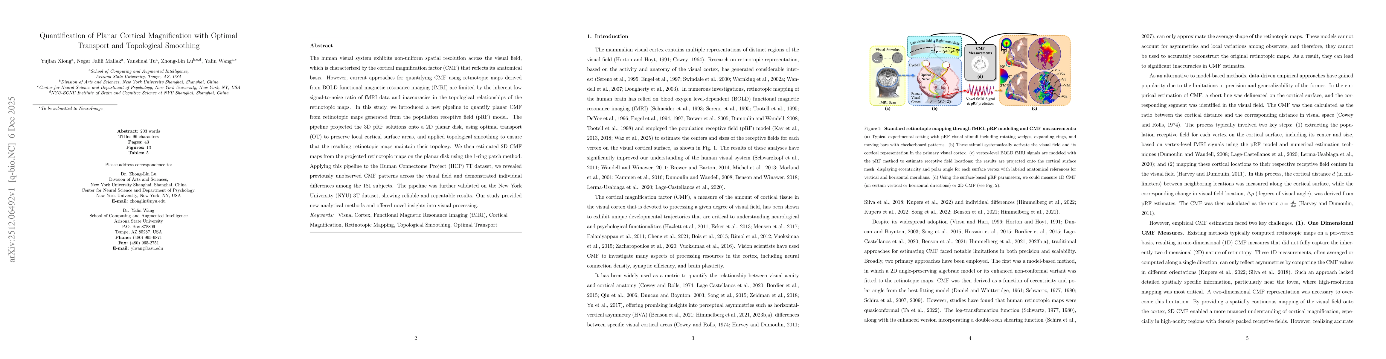
The human visual system exhibits non-uniform spatial resolution across the visual field, which is characterized by the cortical magnification factor (CMF) that reflects its anatomical basis. However, current approaches for quantifying CMF using retinotopic maps derived from BOLD functional magnetic resonance imaging (fMRI) are limited by the inherent low signal-to-noise ratio of fMRI data and inaccuracies in the topological relationships of the retinotopic maps. In this study, we introduced a new pipeline to quantify planar CMF from retinotopic maps generated from the population receptive field (pRF) model. The pipeline projected the 3D pRF solutions onto a 2D planar disk, using optimal transport (OT) to preserve local cortical surface areas, and applied topological smoothing to ensure that the resulting retinotopic maps maintain their topology. We then estimated 2D CMF maps from the projected retinotopic maps on the planar disk using the 1-ring patch method. Applying this pipeline to the Human Connectome Project (HCP) 7T dataset, we revealed previously unobserved CMF patterns across the visual field and demonstrated individual differences among the 181 subjects. The pipeline was further validated on the New York University (NYU) 3T dataset, showing reliable and repeatable results. Our study provided new analytical methods and offered novel insights into visual processing.
AI Key Findings
Get AI-generated insights about this paper's methodology, results, significance, and more — seven facets brought into focus.
Discussion 0