Publication
Metrics
AI Quick Summary
This study employs immunohistochemistry, multiphoton microscopy, and machine learning to quantify the regional organization of smooth muscle bundles in the rat bladder. A Random Forest classifier automates the segmentation of bundles, and the CT-FIRE package determines their orientation, providing essential data for biomechanical bladder models.
Paper Preview
Abstract
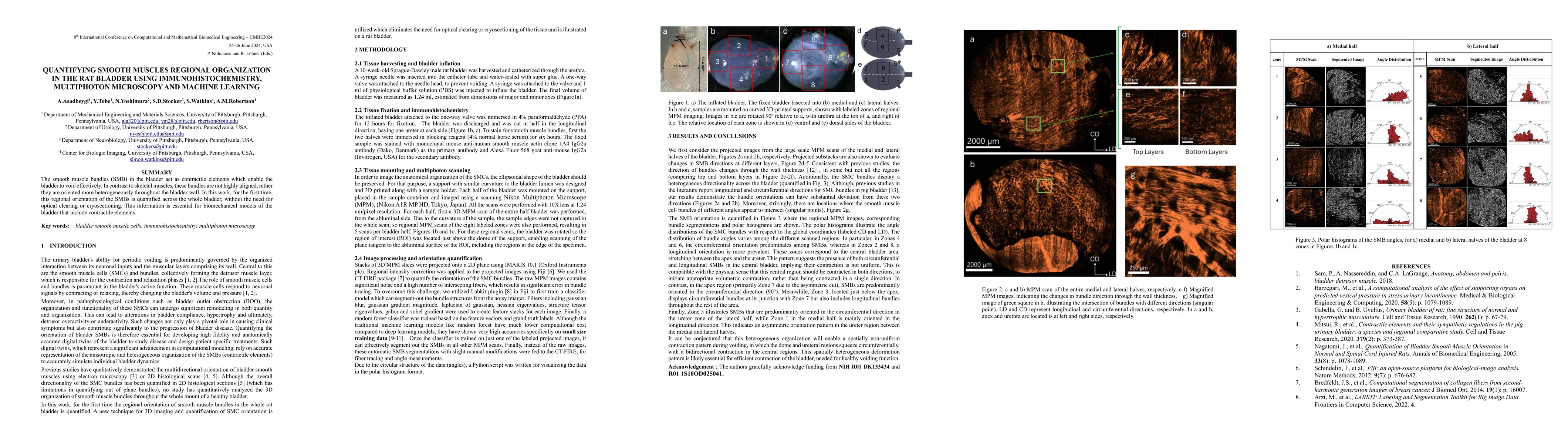
The smooth muscle bundles (SMBs) in the bladder act as contractile elements which enable the bladder to void effectively. In contrast to skeletal muscles, these bundles are not highly aligned, rather they are oriented more heterogeneously throughout the bladder wall. In this work, for the first time, this regional orientation of the SMBs is quantified across the whole bladder, without the need for optical clearing or cryosectioning. Immunohistochemistry staining was utilized to visualize smooth muscle cell actin in multiphoton microscopy (MPM) images of bladder smooth muscle bundles (SMBs). Feature vectors for each pixel were generated using a range of filters, including Gaussian blur, Gaussian gradient magnitude, Laplacian of Gaussian, Hessian eigenvalues, structure tensor eigenvalues, Gabor, and Sobel gradients. A Random Forest classifier was subsequently trained to automate the segmentation of SMBs in the MPM images. Finally, the orientation of SMBs in each bladder region was quantified using the CT-FIRE package. This information is essential for biomechanical models of the bladder that include contractile elements.
AI Key Findings
Get AI-generated insights about this paper's methodology, results, significance, and more — seven facets brought into focus.
Impact
Paper Details
Authors
PDF Preview
Key Terms
Citation Network
Current paper (gray), citations (green), references (blue)
Display is limited for performance on very large graphs.
Discussion 0