Publication

Published: Apr 08, 2026
Categories: q-bio.QM, stat.AP

Metrics

Source: ArXiv

Quick Actions

View PDF

Quick Answers

What methodology did the authors use?

Open-source computational pipeline combining full-field displacement tracking, strain reconstruction, spatial registration, dimensionality reduction (PCA, UMAP, t‑SNE), and topology-based vector-field analysis to extract 16 interpretable metrics from brightfield time‑lapse videos of 670 human iPSC‑derived cardiac microbundles across 20 experimental conditions. More in Methodology →

What are the key results?

Continuous variation of contractile phenotypes across conditions, with intra‑condition variability often exceeding inter‑condition differences. — Principal component analysis reveals a dominant global isotropic contraction mode accounting for ~85% of variance, while higher‑order PCs capture localized saddle‑type deformations present in ~50% of samples. More in Key Results →

Why is this work significant?

The pipeline provides a reproducible, scalable, and interpretable framework for quantifying dynamic cardiac tissue mechanics, enabling cross‑platform comparison and informing computational modeling of engineered myocardium. More in Significance →

What are the main limitations?

High intra‑condition variability limits the ability to detect discrete phenotypic clusters. — Effect sizes in multivariate comparisons are very small, indicating subtle differences between conditions. More in Limitations →

Paper Preview

Abstract

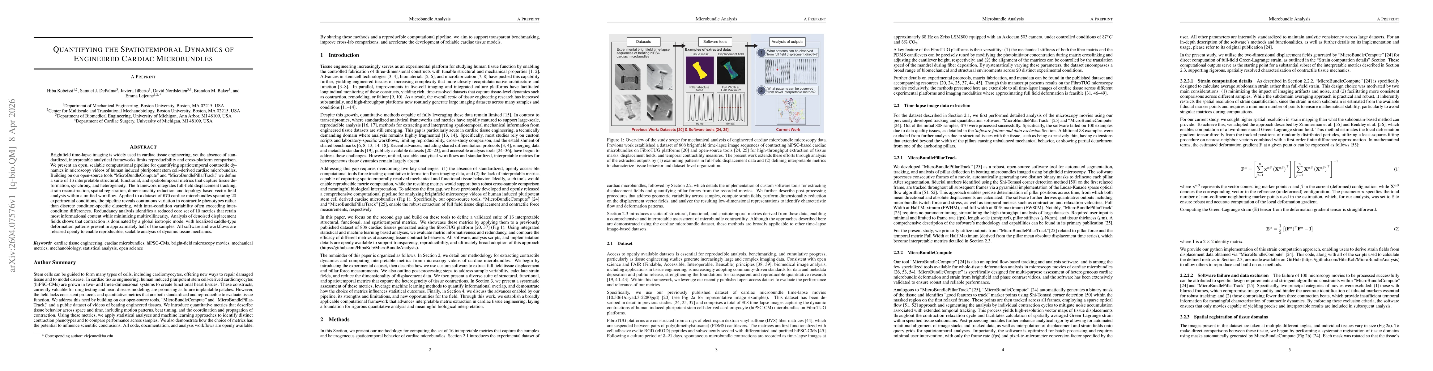
Brightfield time-lapse imaging is widely used in cardiac tissue engineering, yet the absence of standardized, interpretable analytical frameworks limits reproducibility and cross-platform comparison. We present an open, scalable computational pipeline for quantifying spatiotemporal contractile dynamics in microscopy videos of human induced pluripotent stem cell-derived cardiac microbundles. Building on our open-source tools "MicroBundleCompute" and "MicroBundlePillarTrack," we define a suite of 16 interpretable structural, functional, and spatiotemporal metrics that capture tissue deformation, synchrony, and heterogeneity. The framework integrates full-field displacement tracking, strain reconstruction, spatial registration, dimensionality reduction, and topology-based vector-field analysis within a unified workflow. Applied to a dataset of 670 cardiac microbundles spanning 20 experimental conditions, the pipeline reveals continuous variation in contractile phenotypes rather than discrete condition-specific clustering, with intra-condition variability often exceeding inter-condition differences. Redundancy analysis identifies a reduced core set of 10 metrics that retain most informational content while minimizing multicollinearity. Analysis of denoised displacement fields shows that contraction is dominated by a global isotropic mode, with localized saddle-type deformation patterns present in approximately half of the samples. All software and workflows are released openly to enable reproducible, scalable analysis of dynamic tissue mechanics.

AI Key Findings

Generated Apr 11, 2026

Methodology — What approach did the authors take?

Open-source computational pipeline combining full-field displacement tracking, strain reconstruction, spatial registration, dimensionality reduction (PCA, UMAP, t‑SNE), and topology-based vector-field analysis to extract 16 interpretable metrics from brightfield time‑lapse videos of 670 human iPSC‑derived cardiac microbundles across 20 experimental conditions.

Key Results — What are the main findings?

  • Continuous variation of contractile phenotypes across conditions, with intra‑condition variability often exceeding inter‑condition differences.
  • Principal component analysis reveals a dominant global isotropic contraction mode accounting for ~85% of variance, while higher‑order PCs capture localized saddle‑type deformations present in ~50% of samples.
  • Redundancy analysis reduces the metric set to 10 core features that retain most informational content, and multivariate PERMANOVA shows statistically significant but very small effect sizes between conditions.
  • Critical‑point analysis identifies saddle‑point patterns in exactly half of the samples, with prevalence varying by experimental condition.
  • Strong positive correlations between tissue displacement and pillar force confirm that isotropic contraction drives higher contractile force.

Significance — Why does this research matter?

The pipeline provides a reproducible, scalable, and interpretable framework for quantifying dynamic cardiac tissue mechanics, enabling cross‑platform comparison and informing computational modeling of engineered myocardium.

Technical Contribution — What is the technical contribution?

An open‑source, end‑to‑end workflow that unifies displacement tracking, strain reconstruction, and topology‑based vector‑field analysis, producing 16 interpretable metrics and a reduced core set for robust characterization of cardiac microbundle dynamics.

Novelty — What is new about this work?

First comprehensive, interpretable metric suite for engineered cardiac microbundles that demonstrates continuous phenotypic variation rather than discrete clustering, combining topological analysis with PCA and providing openly available tools for reproducible, scalable tissue mechanics analysis.

Limitations — What are the limitations of this study?

  • High intra‑condition variability limits the ability to detect discrete phenotypic clusters.
  • Effect sizes in multivariate comparisons are very small, indicating subtle differences between conditions.
  • The dataset is limited to 20 experimental conditions and 670 samples, which may constrain generalizability.
  • Potential analytical flexibility could inflate false positives if not transparently reported.

Future Work — What did the authors propose for future work?

  • Integrate multimodal data (gene expression, immunostaining) to link mechanical phenotypes with cellular composition.
  • Extend PCA to spatiotemporal modes and develop critical‑point tracking over contraction cycles.
  • Apply larger, more diverse datasets and advanced machine‑learning to refine the minimal metric set.
  • Use the framework to validate and calibrate computational models of cardiac microbundle mechanics.

Paper Details

Paper ID: 2604.07576
Comments: 37 pages, 13 main figures, 3 supplementary figures
Categories:
q-bio.QM stat.AP

How to Cite This Paper

BibTeX
@article{baker2026quantifying,
  title         = {Quantifying the Spatiotemporal Dynamics of Engineered Cardiac Microbundles},
  author        = {Baker, Brendon M. and Nordsletten, David and Lejeune, Emma and others},
  year          = {2026},
  eprint        = {2604.07576},
  archivePrefix = {arXiv},
  primaryClass  = {q-bio.QM},
}
APA 7
Baker, B., Nordsletten, D., Lejeune, E., Kobeissi, H., Jilberto, J., & DePalma, S. (2026). Quantifying the Spatiotemporal Dynamics of Engineered Cardiac Microbundles. arXiv. https://arxiv.org/abs/2604.07576
MLA 9
Baker, Brendon M., et al. "Quantifying the Spatiotemporal Dynamics of Engineered Cardiac Microbundles." arXiv, 2026, arxiv.org/abs/2604.07576.

Comments (0)

This website uses cookies to ensure you get the best experience. Learn more