ArxivLens

Quick Summary:

This paper introduces a novel method for quantifying the x-ray dark-field signal in single-grid imaging, showing that the signal strength correlates to the number of microstructures in the sample. The derived relationship, $2.19\sqrt{N}$, provides a practical approach for quantitative dark-field imaging under limited exposure conditions.

DOI: 10.1364/OE.451834
arXiv Id: 2112.13968
Comments: 20 pages, 9 figures
Date Published: 2022-04-06
Date Updated: 2022-04-06

Categories:

Key Terms (TF-IDF):

imaging (0.281)
dark (0.274)
imaging (0.271)
dark (0.267)
sample (0.244)
sample (0.226)
grid (0.215)
grid (0.208)
sqrt (0.198)
ray (0.196)
ray (0.193)
sqrt (0.188)
signal (0.170)
field (0.166)
signal (0.165)
field (0.162)
microstructure (0.157)
dose (0.156)
exposure (0.153)
unresolved (0.151)
License: http://creativecommons.org/licenses/by/4.0/

Summary:

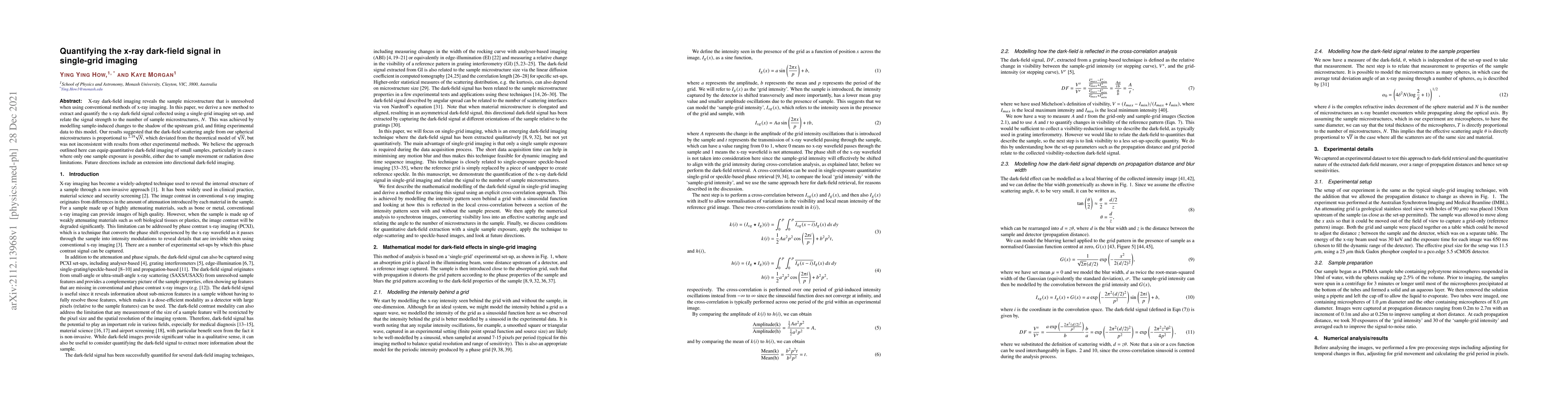
X-ray dark-field imaging reveals the sample microstructure that is unresolved when using conventional methods of x-ray imaging. In this paper, we derive a new method to extract and quantify the x-ray dark-field signal collected using a single-grid imaging set-up, and relate the signal strength to the number of sample microstructures, $N$. This was achieved by modelling sample-induced changes to the shadow of the upstream grid, and fitting experimental data to this model. Our results suggested that the dark-field scattering angle from our spherical microstructures is proportional to $^{2.19}\sqrt{N}$, which deviated from the theoretical model of $\sqrt{N}$, but was not inconsistent with results from other experimental methods. We believe the approach outlined here can equip quantitative dark-field imaging of small samples, particularly in cases where only one sample exposure is possible, either due to sample movement or radiation dose limitations. Future directions include an extension into directional dark-field imaging.

Citation & Reference Network Visualization

This graph shows how this paper is connected to other research. The current paper is shown in dark gray, papers that cite it are in green, and papers it references are in blue. You can:

Current Paper
Papers citing this work (Citations)
Papers cited by this work (References)

• Hover over any node to see the paper title

• Click a node to view that paper's details

• Drag nodes to rearrange the graph

• Use the zoom controls (bottom right) to zoom in/out

Citations and References

Title Authors Year Actions
Hybrid dark-field and attenuation contrast retrieval for laboratory-based X-ray tomography
A. Doherty, I. Buchanan, O. Roche i Morgó, A. Astolfo, S. Savvidis, M. F. M. Gerli, A. Citro, A. Olivo, M. Endrizzi
2024
View Paper
Separating edges from microstructure in X-ray dark-field imaging: evolving and devolving perspectives via the X-ray Fokker-Planck equation.
S. J. Alloo, D. Paganin, M. Croughan, Jannis N. Ahlers, Konstantin M. Pavlov, K. Morgan
2024
View Paper
Grating-based spatial harmonic frequency X-ray imaging for quantitative characterization of structures with sub-detector spatial resolution
M. Gobo, M. G. Hönnicke, M.E. Poletti
2024
View Paper
Correcting directional dark field x-ray imaging artefacts using position dependent image deblurring and attenuation removal
M. Croughan, D. Paganin, S. J. Alloo, Jannis N. Ahlers, Y. Y. How, Stephanie A. Harker, K. Morgan
2024
View Paper
X-ray dark-field via spectral propagation-based imaging
Jannis N. Ahlers, K. M. Pavlov, M. Kitchen, K. Morgan
2023
View Paper
Multimodal intrinsic speckle-tracking (MIST) to extract images of rapidly-varying diffuse X-ray dark-field
S. J. Alloo, K. Morgan, D. Paganin, K. Pavlov
2023
View Paper
A fast implicit X-ray diffusive-dark-field retrieval method using a single mask and exposure
M. Beltran, D. Paganin, M. Croughan, K. Morgan
2023
View Paper
Multimodal Intrinsic Speckle-Tracking (MIST) to extract rapidly-varying diffuse X-ray scatter
S. J. Alloo, K. Morgan, D. Paganin, K. Pavlov
2023
View Paper
Paraxial diffusion-field retrieval
D. Paganin, D. Pelliccia, K. Morgan
2023
View Paper
On the quantification of sample microstructure using single-exposure x-ray dark-field imaging via a single-grid setup
Y. Y. How, D. Paganin, K. Morgan
2022
View Paper
Directional dark-field retrieval with single-grid x-ray imaging.
M. Croughan, Y. Y. How, Allan Pennings, K. Morgan
2022
View Paper
Fast implicit diffusive dark-field retrieval for single-exposure, single-mask x-ray imaging
M. Beltran, D. Paganin, M. Croughan, K. Morgan
2022
View Paper
X-Ray Dark-Field and Phase Retrieval Without Optics, via the Fokker–Planck Equation
Thomas A. Leatham, D. Paganin, K. Morgan
2021
View Paper

Similar Papers

Directional dark-field retrieval with single-grid x-ray imaging

Michelle K Croughan, Ying Ying How, Kaye S Morgan, Allan Pennings

Published: 2023-04-07

In vivo 4D x-ray dark-field lung imaging in mice

David M. Paganin, Ying Ying How, Kaye S. Morgan, Jannis N. Ahlers, Marcus J. Kitchen, Nicole Reyne, Michelle K. Croughan, Patricia Cmielewski, Daniel Batey, Lucy F. Costello, Ronan Smith, Marian Cholewa, Magdalena Kolodziej, Julia Duerr, Marcus A. Mall, Marie-Liesse Asselin-Labat, Martin Donnelley

Published: 2024-11-22

X-ray dark-field via spectral propagation-based imaging

Kaye S. Morgan, Jannis N. Ahlers, Konstantin M. Pavlov, Marcus J. Kitchen

Published: 2023-09-29

Comments (0)

This website uses cookies to ensure you get the best experience. Learn more