Quantifying U-Net Uncertainty in Multi-Parametric MRI-based Glioma Segmentation by Spherical Image Projection
Publication
Metrics
AI Quick Summary
The paper proposes a spherical projection-based U-Net (SPU-Net) for multi-parametric MRI segmentation of gliomas, which generates multiple segmentation predictions and visualizes uncertainty. The SPU-Net model outperformed traditional U-Net and LSU-Net models in both segmentation accuracy and uncertainty quantification, achieving higher Dice coefficients and better uncertainty scores.
Paper Preview
Abstract
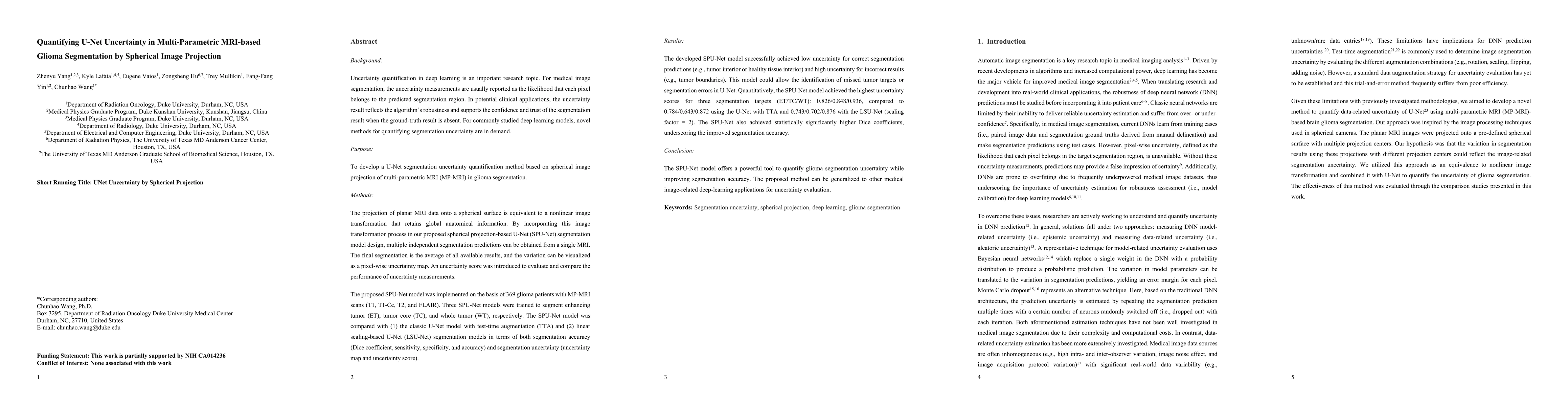
The projection of planar MRI data onto a spherical surface is equivalent to a nonlinear image transformation that retains global anatomical information. By incorporating this image transformation process in our proposed spherical projection-based U-Net (SPU-Net) segmentation model design, multiple independent segmentation predictions can be obtained from a single MRI. The final segmentation is the average of all available results, and the variation can be visualized as a pixel-wise uncertainty map. An uncertainty score was introduced to evaluate and compare the performance of uncertainty measurements. The proposed SPU-Net model was implemented on the basis of 369 glioma patients with MP-MRI scans (T1, T1-Ce, T2, and FLAIR). Three SPU-Net models were trained to segment enhancing tumor (ET), tumor core (TC), and whole tumor (WT), respectively. The SPU-Net model was compared with (1) the classic U-Net model with test-time augmentation (TTA) and (2) linear scaling-based U-Net (LSU-Net) segmentation models in terms of both segmentation accuracy (Dice coefficient, sensitivity, specificity, and accuracy) and segmentation uncertainty (uncertainty map and uncertainty score). The developed SPU-Net model successfully achieved low uncertainty for correct segmentation predictions (e.g., tumor interior or healthy tissue interior) and high uncertainty for incorrect results (e.g., tumor boundaries). This model could allow the identification of missed tumor targets or segmentation errors in U-Net. Quantitatively, the SPU-Net model achieved the highest uncertainty scores for three segmentation targets (ET/TC/WT): 0.826/0.848/0.936, compared to 0.784/0.643/0.872 using the U-Net with TTA and 0.743/0.702/0.876 with the LSU-Net (scaling factor = 2). The SPU-Net also achieved statistically significantly higher Dice coefficients, underscoring the improved segmentation accuracy.
AI Key Findings
Get AI-generated insights about this paper's methodology, results, significance, and more — seven facets brought into focus.
Impact
Paper Details
Authors
PDF Preview
Key Terms
Citation Network
Current paper (gray), citations (green), references (blue)
Display is limited for performance on very large graphs.
Discussion 0