Publication
Metrics
AI Quick Summary
This paper presents a lateral-shearing digital holographic microscope using a two-beam interference technique to quantitatively analyze the morphology of human red blood cells. The system enhances resolution by filtering object information and maintaining fringe contrast, providing 3-D profile and depth information.
Paper Preview
Abstract
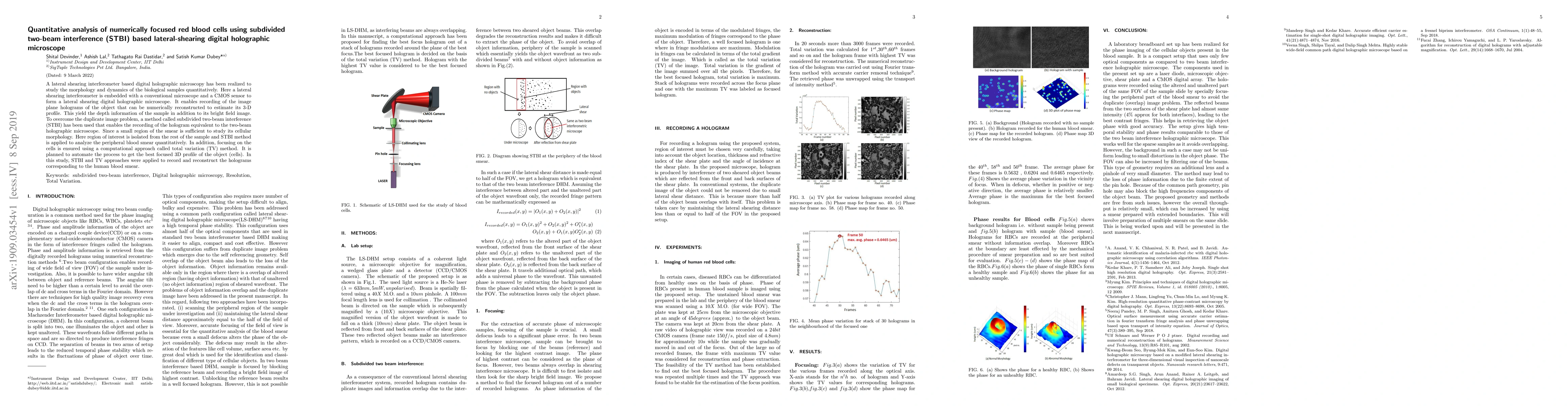
A lateral shear interferometer based digital holographic microscopy has been realized to study the morphology dynamics of Human red blood cells quantitatively. Here, a lateral shear interferometer is embedded with a conventional microscope with a CMOS sensor to form a lateral-shearing digital holographic microscope. It enables recording of image plane holograms of objects that can be numerically reconstructed to estimate its3-D prole. This yields the depth information of the sample in addition to its bright field image. The problem of the duplicate image is addressed by filtering object information from one of the beams, without losing the contrast in the fringes. The contrast in the fringes is maintained by increased reflectivity of the back surface of the shear plate. This improves the resolution in the proposed system.
AI Key Findings
Get AI-generated insights about this paper's methodology, results, significance, and more — seven facets brought into focus.
Impact
Paper Details
Authors
PDF Preview
Key Terms
Citation Network
Current paper (gray), citations (green), references (blue)
Display is limited for performance on very large graphs.
Discussion 0