Quantitative analysis of shadow X-ray Magnetic Circular Dichroism Photo-Emission Electron Microscopy
Publication
Metrics
AI Quick Summary
Researchers developed a method to simulate contrast in Shadow X-ray Magnetic Circular Dichroism Photo-Emission Electron Microscopy (XMCD-PEEM) for quantitative analysis of magnetization texture inside objects.
Paper Preview
Abstract
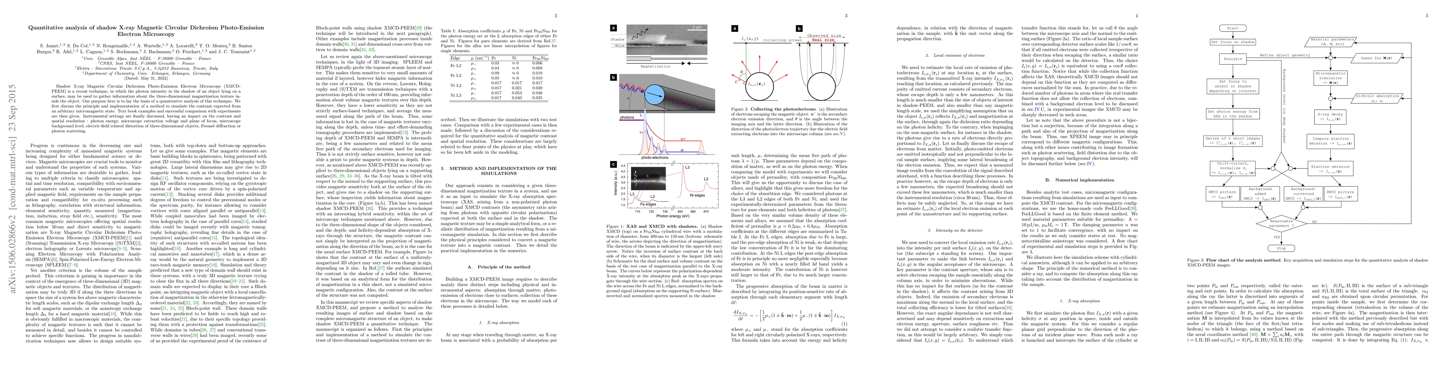
Shadow X-ray Magnetic Circular Dichroism Photo-Emission Electron Microscopy (XMCD-PEEM) is a recent technique, in which the photon intensity in the shadow of an object lying on a surface, may be used to gather information about the three-dimensional magnetization texture inside the object. Our purpose here is to lay the basis of a quantitative analysis of this technique. We first discuss the principle and implementation of a method to simulate the contrast expected from an arbitrary micromagnetic state. Text book examples and successful comparison with experiments are then given. Instrumental settings are finally discussed, having an impact on the contrast and spatial resolution : photon energy, microscope extraction voltage and plane of focus, microscope background level, electric-field related distortion of three-dimensional objects, Fresnel diffraction or photon scattering.
AI Key Findings
Get AI-generated insights about this paper's methodology, results, significance, and more — seven facets brought into focus.
Impact
Paper Details
PDF Preview
Key Terms
Citation Network
Current paper (gray), citations (green), references (blue)
Display is limited for performance on very large graphs.
Discussion 0