Quantitative characterization of the imaging limits of diffuse low-grade oligodendrogliomas
Publication
Metrics
Paper Preview
Abstract
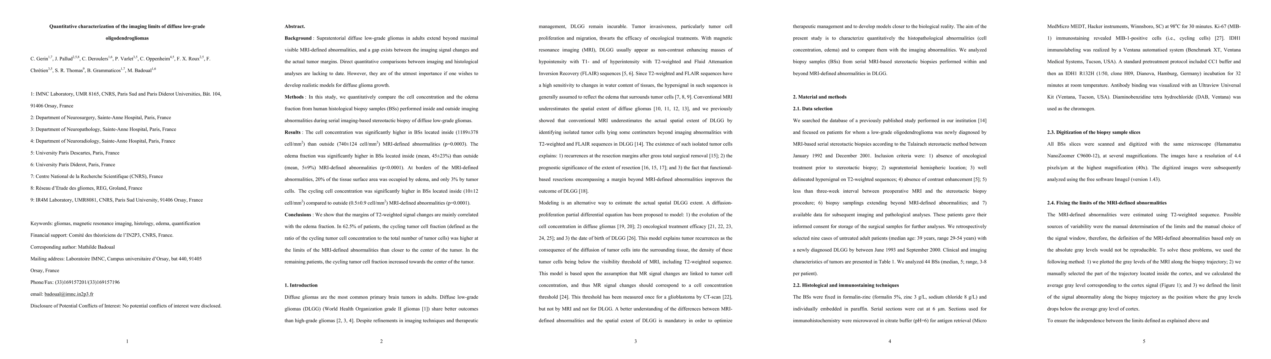
Background : Supratentorial diffuse low-grade gliomas in adults extend beyond maximal visible MRI-defined abnormalities, and a gap exists between the imaging signal changes and the actual tumor margins. Direct quantitative comparisons between imaging and histological analyses are lacking to date. However, they are of the utmost importance if one wishes to develop realistic models for diffuse glioma growth. Methods : In this study, we quantitatively compare the cell concentration and the edema fraction from human histological biopsy samples (BSs) performed inside and outside imaging abnormalities during serial imaging-based stereotactic biopsy of diffuse low-grade gliomas. Results : The cell concentration was significantly higher in BSs located inside (1189 $\pm$ 378 cell/mm$^2$) than outside (740 $\pm$ 124 cell/mm$^2$) MRI-defined abnormalities (p=0.0003). The edema fraction was significantly higher in BSs located inside (mean, 45 $\pm$ 23%) than outside (mean, 5 $\pm$ 9%) MRI-defined abnormalities (p<0.0001). At borders of the MRI-defined abnormalities, 20% of the tissue surface area was occupied by edema, and only 3% by tumor cells. The cycling cell concentration was significantly higher in BSs located inside (10 $\pm$ 12 cell/mm$^2$) compared to outside (0.5 $\pm$ 0.9 cell/mm$^2$) MRI-defined abnormalities (p=0.0001). Conclusions : We show that the margins of T2-weighted signal changes are mainly correlated with the edema fraction. In 62.5% of patients, the cycling tumor cell fraction (defined as the ratio of the cycling tumor cell concentration to the total number of tumor cells) was higher at the limits of the MRI-defined abnormalities than closer to the center of the tumor. In the remaining patients, the cycling tumor cell fraction increased towards the center of the tumor.
AI Key Findings
Get AI-generated insights about this paper's methodology, results, significance, and more — seven facets brought into focus.
Impact
Paper Details
PDF Preview
Key Terms
Citation Network
Current paper (gray), citations (green), references (blue)
Display is limited for performance on very large graphs.
Discussion 0