Quantitative Histopathology of Stained Tissues using Color Spatial Light Interference Microscopy (cSLIM)
Publication
Metrics
AI Quick Summary
This paper introduces color spatial light interference microscopy (cSLIM) as a new quantitative histopathology tool that combines interferometric imaging with color detection, providing both phase maps and standard bright-field images. The study demonstrates cSLIM's potential to offer both diagnostic and prognostic insights from stained tissue samples, addressing the limitations of previous quantitative phase imaging methods reliant on unstained tissues.
Paper Preview
Abstract
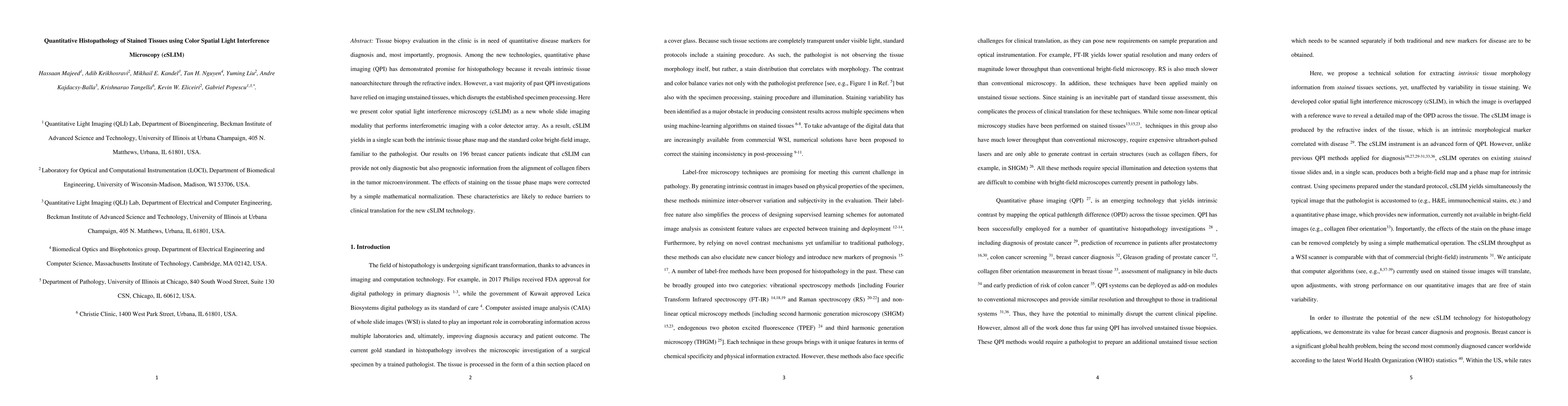
Tissue biopsy evaluation in the clinic is in need of quantitative disease markers for diagnosis and, most importantly, prognosis. Among the new technologies, quantitative phase imaging (QPI) has demonstrated promise for histopathology because it reveals intrinsic tissue nanoarchitecture through the refractive index. However, a vast majority of past QPI investigations have relied on imaging unstained tissues, which disrupts the established specimen processing. Here we present color spatial light interference microscopy (cSLIM) as a new whole slide imaging modality that performs interferometric imaging with a color detector array. As a result, cSLIM yields in a single scan both the intrinsic tissue phase map and the standard color bright-field image, familiar to the pathologist. Our results on 196 breast cancer patients indicate that cSLIM can provide not only diagnostic but also prognostic information from the alignment of collagen fibers in the tumor microenvironment. The effects of staining on the tissue phase maps were corrected by a simple mathematical normalization. These characteristics are likely to reduce barriers to clinical translation for the new cSLIM technology.
AI Key Findings
Get AI-generated insights about this paper's methodology, results, significance, and more — seven facets brought into focus.
Impact
Paper Details
PDF Preview
Key Terms
Citation Network
Current paper (gray), citations (green), references (blue)
Display is limited for performance on very large graphs.
Discussion 0