Quantitative multi-metabolite imaging of Parkinson's disease using AI boosted molecular MRI
Publication
Metrics
Paper Preview
Abstract
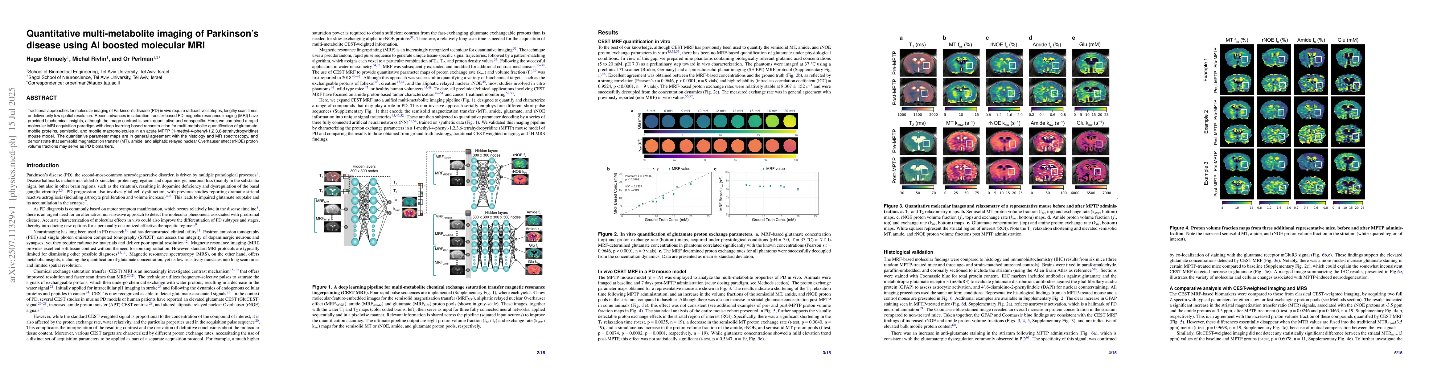
Traditional approaches for molecular imaging of Parkinson's disease (PD) in vivo require radioactive isotopes, lengthy scan times, or deliver only low spatial resolution. Recent advances in saturation transfer-based PD magnetic resonance imaging (MRI) have provided biochemical insights, although the image contrast is semi-quantitative and nonspecific. Here, we combined a rapid molecular MRI acquisition paradigm with deep learning based reconstruction for multi-metabolite quantification of glutamate, mobile proteins, semisolid, and mobile macromolecules in an acute MPTP (1-methyl-4-phenyl-1,2,3,6-tetrahydropyridine) mouse model. The quantitative parameter maps are in general agreement with the histology and MR spectroscopy, and demonstrate that semisolid magnetization transfer (MT), amide, and aliphatic relayed nuclear Overhauser effect (rNOE) proton volume fractions may serve as PD biomarkers.
AI Key Findings
Get AI-generated insights about this paper's methodology, results, significance, and more — seven facets brought into focus.
Impact
Paper Details
Authors
PDF Preview
Citation Network
Current paper (gray), citations (green), references (blue)
Display is limited for performance on very large graphs.
Discussion 0