Publication
Metrics
AI Quick Summary
This paper presents the first quantitative X-ray phase-contrast microtomogram using a compact laser-driven betatron source, demonstrating its potential to bridge the performance gap between synchrotron and conventional X-ray sources. The study highlights the excellent spatial coherence and spectral stability of betatron radiation for advanced biomedical imaging applications.
Paper Preview
Abstract
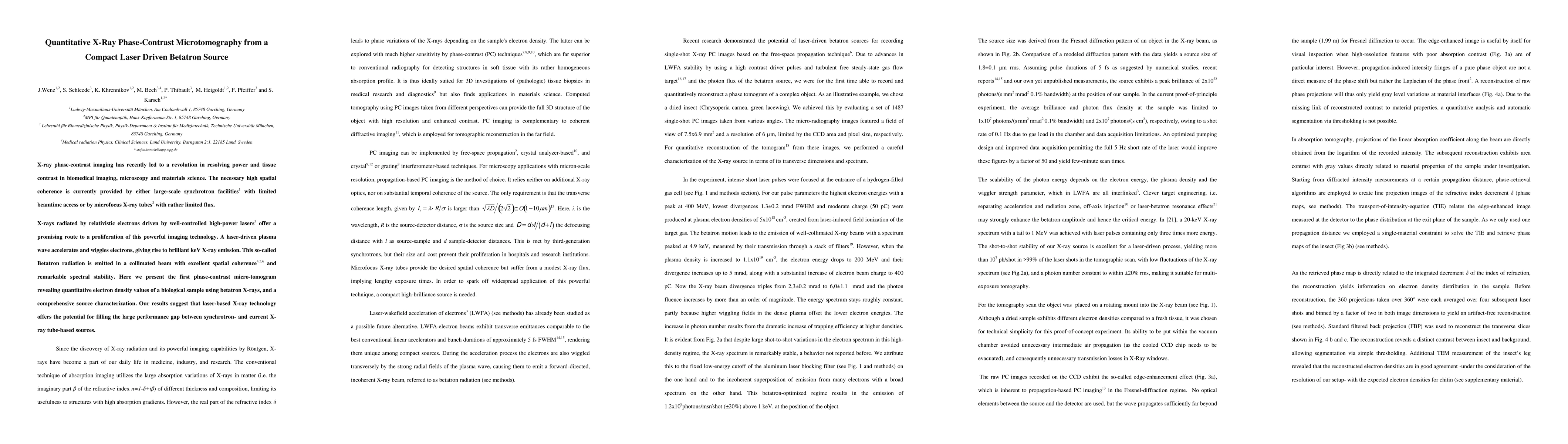
X-ray phase-contrast imaging has recently led to a revolution in resolving power and tissue contrast in biomedical imaging, microscopy and materials science. The necessary high spatial coherence is currently provided by either large-scale synchrotron facilities with limited beamtime access or by microfocus X-ray tubes with rather limited flux. X-rays radiated by relativistic electrons driven by well-controlled high-power lasers offer a promising route to a proliferation of this powerful imaging technology. A laser-driven plasma wave accelerates and wiggles electrons, giving rise to brilliant keV X-ray emission. This so-called Betatron radiation is emitted in a collimated beam with excellent spatial coherence and remarkable spectral stability. Here we present the first phase-contrast micro-tomogram revealing quantitative electron density values of a biological sample using betatron X-rays, and a comprehensive source characterization. Our results suggest that laser-based X-ray technology offers the potential for filling the large performance gap between synchrotron- and current X-ray tube-based sources.
AI Key Findings
Get AI-generated insights about this paper's methodology, results, significance, and more — seven facets brought into focus.
Impact
Paper Details
PDF Preview
Key Terms
Citation Network
Current paper (gray), citations (green), references (blue)
Display is limited for performance on very large graphs.
Discussion 0