Quantum optical coherence tomography of a biological sample
Publication
Metrics
AI Quick Summary
This paper demonstrates the first experimental application of quantum optical coherence tomography (QOCT) using an entangled-photon light source to achieve dispersion-immune axial optical sectioning in a biological sample. The study presents 3D images of onion-skin tissue coated with gold nanoparticles in the form of 2D sections from different orientations.
Paper Preview
Abstract
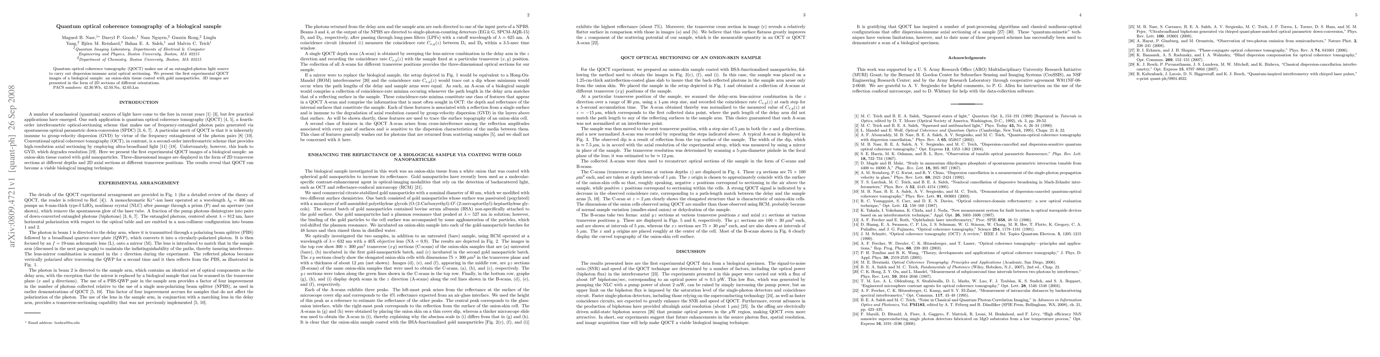
Quantum optical coherence tomography (QOCT) makes use of an entangled-photon light source to carry out dispersion-immune axial optical sectioning. We present the first experimental QOCT images of a biological sample: an onion-skin tissue coated with gold nanoparticles. 3D images are presented in the form of 2D sections of different orientations.
AI Key Findings
Get AI-generated insights about this paper's methodology, results, significance, and more — seven facets brought into focus.
Impact
Paper Details
Authors
PDF Preview
Key Terms
Citation Network
Current paper (gray), citations (green), references (blue)
Display is limited for performance on very large graphs.
Discussion 0