QuickTumorNet is a fast, automated deep learning method for segmenting brain tumors from MRI scans, addressing the limitations of manual segmentation. The model, based on a modified QuickNAT CNN, accurately segments three tumor types, aiding clinicians in diagnosis and treatment.
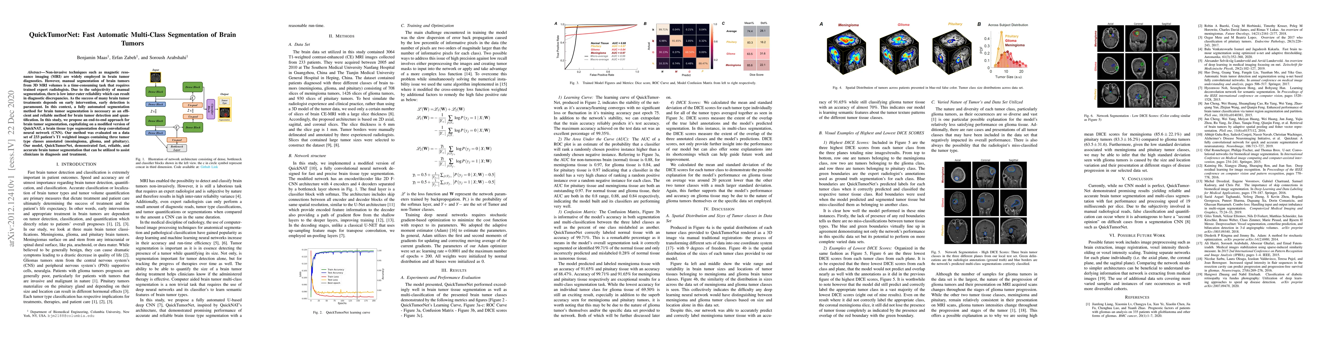
Non-invasive techniques such as magnetic resonance imaging (MRI) are widely employed in brain tumor diagnostics. However, manual segmentation of brain tumors from 3D MRI volumes is a time-consuming task that requires trained expert radiologists. Due to the subjectivity of manual segmentation, there is low inter-rater reliability which can result in diagnostic discrepancies. As the success of many brain tumor treatments depends on early intervention, early detection is paramount. In this context, a fully automated segmentation method for brain tumor segmentation is necessary as an efficient and reliable method for brain tumor detection and quantification. In this study, we propose an end-to-end approach for brain tumor segmentation, capitalizing on a modified version of QuickNAT, a brain tissue type segmentation deep convolutional neural network (CNN). Our method was evaluated on a data set of 233 patient's T1 weighted images containing three tumor type classes annotated (meningioma, glioma, and pituitary). Our model, QuickTumorNet, demonstrated fast, reliable, and accurate brain tumor segmentation that can be utilized to assist clinicians in diagnosis and treatment.
This graph shows how this paper is connected to other research. The current paper is shown in dark gray, papers that cite it are in green, and papers it references are in blue. You can:
• Hover over any node to see the paper title
• Click a node to view that paper's details
• Drag nodes to rearrange the graph
• Use the zoom controls (bottom right) to zoom in/out
Title | Authors | Year | Actions |
---|---|---|---|
Advanced Brain Tumor Segmentation Using UNet and Multiple Residual Networks
|
Ramya D, S. K. Manigandan, J. Velmurugan, A. Vishnukumar, V. UmaRani, P. Reddy
|
2024
|
View Paper |
Brain Disease Detection using PSPNet for Multiclass Segmentation
|
D. Jeni, Jeba Seeli, Reg.No, Dr.K.K.Thanammal
|
2023
|
View Paper |
A novel machine learning approach on texture analysis for automatic breast microcalcification diagnosis classification of mammogram images
|
Zahra Maghsoodzadeh Sarvestani, J. Jamali, M. Taghizadeh, Mohammad Hosein Fatehi Dindarloo
|
2023
|
View Paper |
Efficient segmentation and classification of the tumor using improved encoder-decoder architecture in brain MRI images
|
Archana Ingle, M. Roja, Manoj S. Sankhe, Deepak Patkar
|
2022
|
View Paper |
The relationship between talent management and the efficiency of head nurses and senior and middle managers from the educational and medical centers in Tabriz, Iran – a case study
|
Sajad Hadi Chelan, K. Alimohammadzadeh, A. Maher
|
2022
|
View Paper |
Salvaging tumor from T1-weighted CE-MR images using automatic segmentation techniques
|
Amar Saraswat, Neeta Sharma
|
2022
|
View Paper |
DT-CWT: A New Feature for Tumor Classification in Breast DCE-MRI
|
Parinaz Eskandari, S. B. Shokouhi
|
2021
|
View Paper |
Automated Atlas-Based Segmentation of Breast Region in 3-D Magnetic Resonance Imaging (MRI) Using FCM Method
|
Parinaz Eskandari, S. B. Shokouhi
|
2021
|
View Paper |
ai-corona: Radiologist-assistant deep learning framework for COVID-19 diagnosis in chest CT scans
|
Mehdi Yousefzadeh, P. Esfahanian, Seyed Mohammad Sadegh Movahed, S. Gorgin, D. Rahmati, A. Abedini, S. Nadji, S. Haseli, Mehrdad Bakhshayesh karam, A. Kiani, Meisam Hoseinyazdi, Jafar Roshandel, Reza Lashgari
|
2020
|
View Paper |
Characteristics of Computerized Tomography (CT) Scan in COVID-19 Patients and Their Relationship with Mortality Rate
|
M. Khademalizadeh, F. Mohammadi, A. Zolghadrasli, S. Jafari, S. Roosta, H. Asadi, A. Salimi, H. Bazrafshan, F. Zibaeenejad, S. Mohammadi
|
2022
|
View Paper |
Comments (0)