Radiologic Image-based Statistical Shape Analysis of Brain Tumors
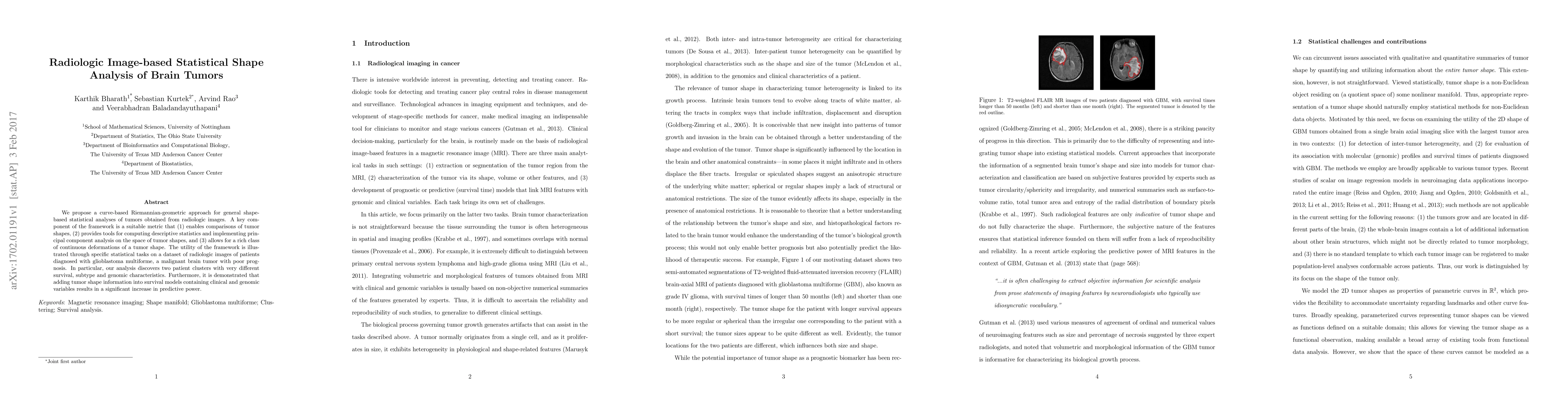
Publication
Metrics
AI Quick Summary
This paper introduces a Riemannian-geometric approach for statistical shape analysis of brain tumors from radiologic images, revealing two distinct patient clusters with different survival, subtype, and genomic characteristics. The method enhances predictive power for survival models when tumor shape information is included alongside clinical and genomic data.
Paper Preview
Abstract
We propose a curve-based Riemannian-geometric approach for general shape-based statistical analyses of tumors obtained from radiologic images. A key component of the framework is a suitable metric that (1) enables comparisons of tumor shapes, (2) provides tools for computing descriptive statistics and implementing principal component analysis on the space of tumor shapes, and (3) allows for a rich class of continuous deformations of a tumor shape. The utility of the framework is illustrated through specific statistical tasks on a dataset of radiologic images of patients diagnosed with glioblastoma multiforme, a malignant brain tumor with poor prognosis. In particular, our analysis discovers two patient clusters with very different survival, subtype and genomic characteristics. Furthermore, it is demonstrated that adding tumor shape information into survival models containing clinical and genomic variables results in a significant increase in predictive power.
AI Key Findings
Get AI-generated insights about this paper's methodology, results, significance, and more — seven facets brought into focus.
Impact
Paper Details
PDF Preview
Key Terms
Citation Network
Current paper (gray), citations (green), references (blue)
Display is limited for performance on very large graphs.
Discussion 0