Publication
Metrics
AI Quick Summary
This study developed a 3D U-Net deep learning model to achieve radiologist-level segmentation of breast cancers on MRI scans, achieving a median Dice score of 0.77, equivalent to radiologists' performance, demonstrating its potential for clinical use in breast cancer detection.
Paper Preview
Abstract
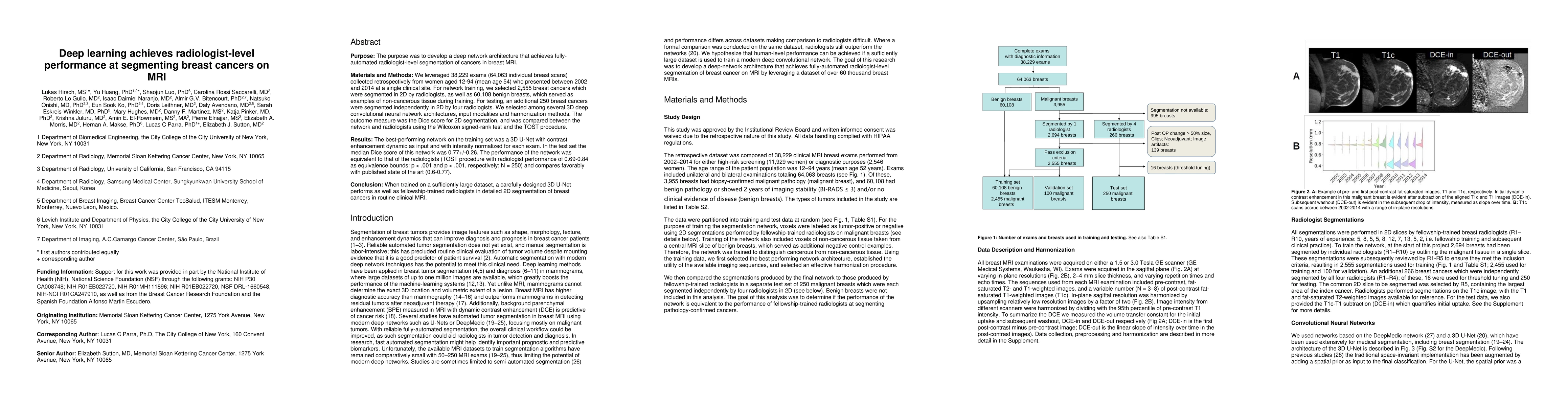
Purpose: To develop a deep network architecture that would achieve fully automated radiologist-level segmentation of cancers at breast MRI. Materials and Methods: In this retrospective study, 38229 examinations (composed of 64063 individual breast scans from 14475 patients) were performed in female patients (age range, 12-94 years; mean age, 52 years +/- 10 [standard deviation]) who presented between 2002 and 2014 at a single clinical site. A total of 2555 breast cancers were selected that had been segmented on two-dimensional (2D) images by radiologists, as well as 60108 benign breasts that served as examples of noncancerous tissue; all these were used for model training. For testing, an additional 250 breast cancers were segmented independently on 2D images by four radiologists. Authors selected among several three-dimensional (3D) deep convolutional neural network architectures, input modalities, and harmonization methods. The outcome measure was the Dice score for 2D segmentation, which was compared between the network and radiologists by using the Wilcoxon signed rank test and the two one-sided test procedure. Results: The highest-performing network on the training set was a 3D U-Net with dynamic contrast-enhanced MRI as input and with intensity normalized for each examination. In the test set, the median Dice score of this network was 0.77 (interquartile range, 0.26). The performance of the network was equivalent to that of the radiologists (two one-sided test procedures with radiologist performance of 0.69-0.84 as equivalence bounds, P <= .001 for both; n = 250). Conclusion: When trained on a sufficiently large dataset, the developed 3D U-Net performed as well as fellowship-trained radiologists in detailed 2D segmentation of breast cancers at routine clinical MRI.
AI Key Findings
Get AI-generated insights about this paper's methodology, results, significance, and more — seven facets brought into focus.
Impact
Paper Details
Authors
PDF Preview
Key Terms
Citation Network
Current paper (gray), citations (green), references (blue)
Display is limited for performance on very large graphs.
Discussion 0