Rapid 2D 23Na MRI of the calf using a denoising convolutional neural network
Publication
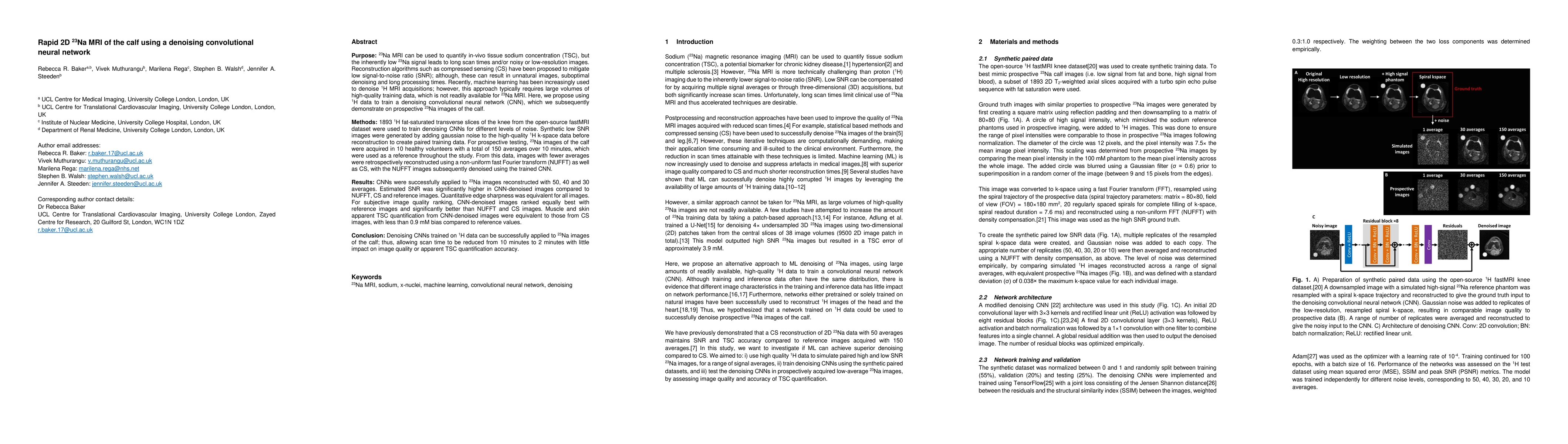
Metrics
AI Quick Summary
This study trains a denoising convolutional neural network (CNN) using 1H MRI data to improve 23Na MRI of the calf, reducing scan times from 10 to 2 minutes. The CNN significantly enhances signal-to-noise ratio and image quality compared to traditional reconstruction methods, while maintaining accurate tissue sodium concentration quantification.
Paper Preview
Abstract
23Na MRI can be used to quantify in-vivo tissue sodium concentration (TSC), but the low 23Na signal leads to long scan times and/or noisy or low-resolution images. Reconstruction algorithms such as CS have been proposed to mitigate low SNR; although, these can result in unnatural images, suboptimal denoising and long processing times. Recently, ML has been used to denoise 1H MRI acquisitions; however, this approach typically requires large volumes of high-quality training data, which is not readily available for 23Na MRI. Here, we train a denoising CNN using 1H data, which we subsequently demonstrate on prospective 23Na images of the calf. 1893 1H transverse slices of the knee were used to train denoising CNNs for different levels of noise. Low SNR images were generated by adding gaussian noise to the high-quality 1H kspace data before reconstruction to create paired training data. For prospective testing, 23Na images of the calf were acquired in 10 volunteers with 150 averages, which were used as a reference throughout the study. From this data, lower-average images were reconstructed using a NUFFT as well as CS, with the NUFFT images subsequently denoised using the trained CNN. CNNs were successfully applied to 23Na images reconstructed with 50, 40 and 30 averages. SNR was significantly higher in CNN images compared to NUFFT, CS and reference images. Edge sharpness was equivalent for all images. For image quality ranking, CNN images ranked equally best with reference images and significantly better than NUFFT and CS images. Muscle and skin TSC quantification from CNN images were equivalent to those from CS images, with <0.9 mM bias compared to reference values. Denoising CNNs trained on 1H data can be successfully applied to 23Na images of the calf; thus, allowing scan time to be reduced from 10 minutes to 2 minutes with little impact on image quality or TSC quantification accuracy.
AI Key Findings
Get AI-generated insights about this paper's methodology, results, significance, and more — seven facets brought into focus.
Impact
Paper Details
Authors
PDF Preview
Key Terms
Citation Network
Current paper (gray), citations (green), references (blue)
Display is limited for performance on very large graphs.
Discussion 0