Quick Summary:

This paper introduces Rapid EBSD, a method that combines forescatter electron imaging with sparse EBSD sampling to efficiently map crystal phase and orientation, enhancing microstructure characterization. It demonstrates the technique on cobalt superalloy and titanium alloy samples, showing its efficiency over conventional EBSD analysis.

Summary

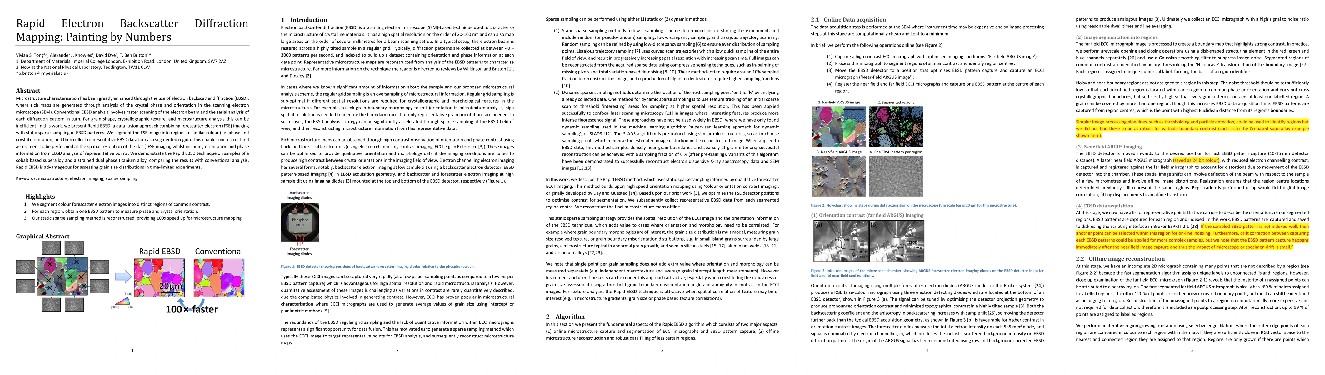
Microstructure characterisation has been greatly enhanced through the use of electron backscatter diffraction (EBSD), where rich maps are generated through analysis of the crystal phase and orientation in the scanning electron microscope (SEM). Conventional EBSD analysis involves raster scanning of the electron beam and the serial analysis of each diffraction pattern in turn. For grain shape, crystallographic texture, and microstructure analysis this can be inefficient. In this work, we present Rapid EBSD, a data fusion approach combining forescatter electron (FSE) imaging with static sparse sampling of EBSD patterns. We segment the FSE image into regions of similar colour (i.e. phase and crystal orientation) and then collect representative EBSD data for each segmented region. This enables microstructural assessment to be performed at the spatial resolution of the (fast) FSE imaging whilst including orientation and phase information from EBSD analysis of representative points. We demonstrate the Rapid EBSD technique on samples of a cobalt based superalloy and a strained dual phase titanium alloy, comparing the results with conventional analysis. Rapid EBSD is advantageous for assessing grain size distributions in time-limited experiments.

AI Key Findings

Get AI-generated insights about this paper's methodology, results, and significance.

Paper Details

PDF Preview

Download PDF

Key Terms

orientation (0.260)
rapid (0.241)
grain (0.234)
scanning (0.224)
representative (0.219)
diffraction (0.219)
electron (0.217)
crystal (0.193)
analysis (0.178)
imaging (0.175)
phase (0.174)
conventional (0.171)
colour (0.145)
collect (0.142)
work present (0.127)
assessment (0.124)
greatly (0.120)
involves (0.118)
turn (0.115)
fusion (0.115)

Citation Network

Current paper (gray), citations (green), references (blue)

Current Paper
Citations
References
Click to view
Title Authors Year Actions
Compressive electron backscatter diffraction imaging
Zoe Broad, Alex W Robinson, J. Wells, D. Nicholls, A. Moshtaghpour, A. Kirkland, Nigel D. Browning
2024
View Paper
Dynamic sampling for scanned microscopy modalities
Zachary Varley, Gregory S. Rohrer, Marc De Graef
2024
View Paper
High‐throughput determination of grain size distributions by EBSD with low‐discrepancy sampling
Timothy J. H. Long, William Holbrook, Todd C. Hufnagel, Tim Mueller
2023
View Paper
The mechanism of Fe-rich intermetallic compound formation and growth on inoculants revealed by electron backscattered diffraction and X-ray imaging
S. Feng, Zelong Jin, W. Du, Insung Han, A. Lui, Xiaoping Zhou, P. Shearing, P. Grant, E. Liotti
2023
View Paper
Q-RBSA: high-resolution 3D EBSD map generation using an efficient quaternion transformer network
Devendra K. Jangid, Neal R. Brodnik, M. Echlin, T. Pollock, S. Daly, B. S. Manjunath
2023
View Paper
Microstructure and crystal structure dependence of microwave dielectric properties of non-stoichiometric (Sr0.7Ca0.3)z(Zr0.95Ti0.05)O3 perovskite ceramics
Qingyang Pang, Yanyan Li, Fan Yang, Zi-Qi Liu, Xin Li, Huarong Cheng, Shuying Sun, Ying Chen, Genshui Wang
2022
View Paper
Development of a new, fully automated system for electron backscatter diffraction (EBSD)-based large volume three-dimensional microstructure mapping using serial sectioning by mechanical polishing, and its application to the analysis of special boundaries in 316L stainless steel.
S. Tsai, P. Konijnenberg, Ivan Gonzalez, Samuel Hartke, Thomas A. Griffiths, M. Herbig, Kaori Kawano-Miyata, A. Taniyama, N. Sano, S. Zaefferer
2022
View Paper
An Acquisition Parameter Study for Machine-Learning-Enabled Electron Backscatter Diffraction
Kevin Kaufmann, K. Vecchio
2021
View Paper
Correction of artefacts associated with large area EBSD.
B. Winiarski, A. Gholinia, K. Mingard, M. Gee, G. Thompson, P. Withers
2021
View Paper
Efficient few-shot machine learning for classification of EBSD patterns
Kevin Kaufmann, Hobson Lane, Xiao Liu, K. Vecchio
2021
View Paper
Adaptive domain misorientation approach for the EBSD measurement of deformation induced dislocation sub-structures.
P. Lehto
2021
View Paper
Characterization of Microstructure in Additively Manufactured 316L using Automated Serial Sectioning
D. Rowenhorst, L. Nguyen, Aeriel D. Murphy-Leonard, R. Fonda
2020
View Paper
Deep Neural Network Enabled Space Group Identification in EBSD
Kevin Kaufmann, Chaoyi Zhu, Alexander S. Rosengarten, K. Vecchio
2020
View Paper
TrueEBSD: Correcting spatial distortions in electron backscatter diffraction maps.
Vivian S. Tong, Thomas Benjamin Britton
2019
View Paper
Advancing characterisation with statistics from correlative electron diffraction and X-ray spectroscopy, in the scanning electron microscope.
TP McAuliffe, A. Foden, C. Bilsland, D. D. Mountanou, D. Dye, T. B. Britton
2019
View Paper

Comments (0)

This website uses cookies to ensure you get the best experience. Learn more