Publication
Metrics
AI Quick Summary
This research introduces SIFA and LipoFRET, two single-molecule imaging methods for observing nanoscale dynamics during membrane protein insertion with sub-nanometer precision, facilitating a deeper understanding of protein-membrane interactions.
Paper Preview
Abstract
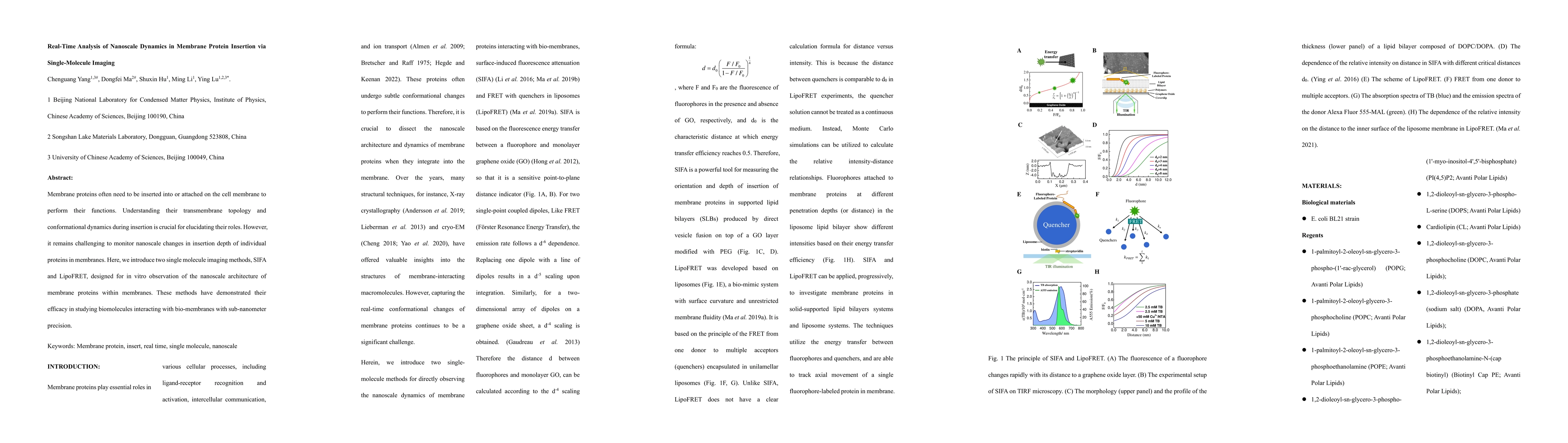
Membrane proteins often need to be inserted into or attached on the cell membrane to perform their functions. Understanding their transmembrane topology and conformational dynamics during insertion is crucial for elucidating their roles. However, it remains challenging to monitor nanoscale changes in insertion depth of individual proteins in membranes. Here, we introduce two single molecule imaging methods, SIFA and LipoFRET, designed for in vitro observation of the nanoscale architecture of membrane proteins within membranes. These methods have demonstrated their efficacy in studying biomolecules interacting with bio-membranes with sub-nanometer precision.
AI Key Findings
Get AI-generated insights about this paper's methodology, results, significance, and more — seven facets brought into focus.
Paper Details
Authors
PDF Preview
Related Papers
No references found for this paper.
Discussion 0