ArxivLens

Quick Summary:

This paper presents a real-time multi-view deconvolution method for light-sheet microscopy that leverages GPU processing to simultaneously fuse and deconvolve 3D images, significantly reducing the time required compared to traditional methods. The approach achieves real-time performance by processing cross-sectional planes individually.

arXiv Id: 1503.07998
Comments: 8 pages, 5 figures, submitted to Bioinformatics
Date Published: 2015-03-30
Date Updated: 2015-03-30

Categories:

Key Terms (TF-IDF):

views (0.332)
views (0.305)
processing (0.283)
processing (0.272)
multi view (0.200)
multiple (0.189)
bottleneck (0.189)
bottleneck (0.188)
gpu (0.186)
sheet (0.185)
individually (0.183)
gpu (0.183)
multiple (0.183)
planes (0.181)
planes (0.181)
acquisition (0.180)
microscopy (0.176)
employs (0.174)
employs (0.171)
acquired (0.170)
License: http://arxiv.org/licenses/nonexclusive-distrib/1.0/

Summary:

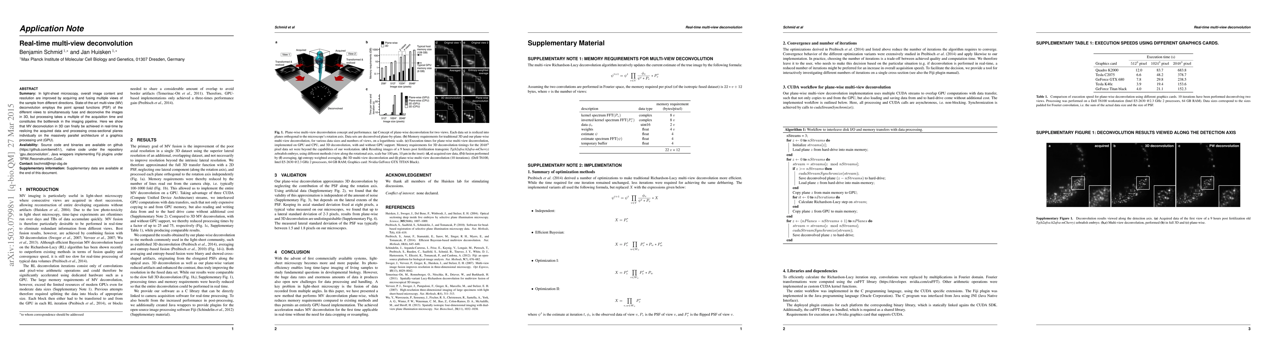
In light-sheet microscopy, overall image content and resolution are improved by acquiring and fusing multiple views of the sample from different directions. State-of-the-art multi-view (MV) deconvolution employs the point spread functions (PSF) of the different views to simultaneously fuse and deconvolve the images in 3D, but processing takes a multiple of the acquisition time and constitutes the bottleneck in the imaging pipeline. Here we show that MV deconvolution in 3D can finally be achieved in real-time by reslicing the acquired data and processing cross-sectional planes individually on the massively parallel architecture of a graphics processing unit (GPU).

Citation & Reference Network Visualization

This graph shows how this paper is connected to other research. The current paper is shown in dark gray, papers that cite it are in green, and papers it references are in blue. You can:

Current Paper
Papers citing this work (Citations)
Papers cited by this work (References)

• Hover over any node to see the paper title

• Click a node to view that paper's details

• Drag nodes to rearrange the graph

• Use the zoom controls (bottom right) to zoom in/out

Citations and References

Title Authors Year Actions
Perspective on Multiview Super‐Resolution Microscopy: Innovations and Future Directions
N. Rabiee
2025
View Paper
Image processing tools for petabyte-scale light sheet microscopy data
Xiongtao Ruan, Matthew Mueller, Gaoxiang Liu, Frederik Görlitz, Tian-Ming Fu, D. Milkie, Joshua L. Lillvis, Alexander Kuhn, Johnny Gan Chong, J. L. Hong, Chu Yi Aaron Herr, Wilmene Hercule, Marc Nienhaus, Alison N. Killilea, Eric Betzig, S. Upadhyayula
2024
View Paper
Image restoration of degraded time-lapse microscopy data mediated by near-infrared imaging
N. Gritti, R. Power, Alyssa L. Graves, J. Huisken
2024
View Paper
Multi-view clustering by CPS-merge analysis with application to multimodal single-cell data
Lixiang Zhang, Lin Lin, Jia Li
2023
View Paper
Cost-effective high-speed, three-dimensional live-cell imaging of HIV-1 transfer at the T cell virological synapse
Alice Sandmeyer, Lili Wang, W. Hübner, Marcel Müller, Benjamin Chen, T. Huser
2022
View Paper
A light sheet fluorescence microscopy protocol for Caenorhabditis elegans larvae and adults
Jayson J. Smith, Isabel W Kenny, Carsten Wolff, R. Cray, Abhishek Kumar, D. Sherwood, D. Matus
2022
View Paper
Two plus one is almost three: a fast approximation for multi-view deconvolution
Manuel Hüpfel, Manuel Fernandez Merino, Johannes Bennemann, Masanari Takamiya, S. Rastegar, A. Tursch, Thomas Holstein, G. Nienhaus
2021
View Paper
Navigating the Light-Sheet Image Analysis Software Landscape: Concepts for Driving Cohesion From Data Acquisition to Analysis
H. Gibbs, S. Mota, N. Hart, Sun Won Min, Alex O. Vernino, Anna L. Pritchard, Anindito Sen, S. Vitha, Sreeja Sarasamma, A. McIntosh, A. Yeh, A. C. Lekven, D. McCreedy, Kristen C. Maitland, Lisa M. Perez
2021
View Paper
Open microscopy in the life sciences: quo vadis?
J. Hohlbein, Benedict Diederich, Barbora Marsíková, E. Reynaud, S. Holden, W. Jahr, R. Haase, Kirti Prakash
2021
View Paper
Three-dimensional bright-field microscopy with isotropic resolution based on multi-view acquisition and image fusion reconstruction
Gianmaria Calisesi, A. Candeo, A. Farina, C. D'Andrea, V. Magni, G. Valentini, A. Pistocchi, Alex Costa, A. Bassi
2020
View Paper
Comparison of Multiscale Imaging Methods for Brain Research
Jessica Tröger, C. Hoischen, B. Perner, S. Monajembashi, A. Barbotin, A. Löschberger, C. Eggeling, M. Kessels, B. Qualmann, P. Hemmerich
2020
View Paper
3D Hessian deconvolution of thick light-sheet z-stacks for high-contrast and high-SNR volumetric imaging
Zhe Zhang, Dongzhou Gou, Fan Feng, Ruyi Zheng, Ke Du, Hongrun Yang, Guangyi Zhang, Huitao Zhang, L. Tao, Liangyi Chen, Heng Mao
2020
View Paper
Optical Aberration Correction via Phase Diversity and Deep Learning
A. Krishnan, C. Belthangady, Clara Nyby, Merlin Lange, Bin Yang, Loic A. Royer
2020
View Paper
Integration of the ImageJ Ecosystem in KNIME Analytics Platform
Christian Dietz, C. Rueden, Stefan Helfrich, E. T. Dobson, Martin Horn, J. Eglinger, Edward L. Evans, Dalton T. Mclean, T. Novitskaya, W. Ricke, N. Sherer, A. Zijlstra, M. Berthold, K. Eliceiri
2020
View Paper
Fast objective coupled planar illumination microscopy
Cody J. Greer, T. Holy
2018
View Paper
Adaptive particle representation of fluorescence microscopy images
B. Cheeseman, Ulrik Günther, Krzysztof Gonciarz, Mateusz Susik, I. Sbalzarini
2018
View Paper
Toward quantitative three-dimensional microvascular networks segmentation with multiview light-sheet fluorescence microscopy
Pol Kennel, L. Teyssedre, J. Colombelli, F. Plouraboué
2018
View Paper
Matrix elasticity regulates mesenchymal stem cell chemotaxis
Neha Saxena, Pankaj Mogha, Silalipi Dash, A. Majumder, S. Jadhav, S. Sen
2018
View Paper
Light-sheet microscopy: a tutorial
Omar E. Olarte, J. Andilla, E. Gualda, P. Loza-Álvarez
2018
View Paper
Forget Pixels: Adaptive Particle Representation of Fluorescence Microscopy Images
B. Cheeseman, Ulrik Günther, Mateusz Susik, Krzysztof Gonciarz, I. Sbalzarini
2018
View Paper
Developing 3D microscopy with CLARITY on human brain tissue: Towards a tool for informing and validating MRI-based histology
M. Morawski, E. Kirilina, Nico Scherf, C. Jäger, K. Reimann, R. Trampel, Filippos Gavriilidis, S. Geyer, B. Biedermann, T. Arendt, N. Weiskopf
2017
View Paper
ReViMS: Software tool for estimating the volumes of 3-D multicellular spheroids imaged using a light sheet fluorescence microscope.
F. Piccinini, A. Tesei, M. Zanoni, A. Bevilacqua
2017
View Paper
Intraoperative histology: Lightning 3D histopathology
R. Power, J. Huisken
2017
View Paper
A guide to light-sheet fluorescence microscopy for multiscale imaging
R. Power, J. Huisken
2017
View Paper
Quantitating the cell: turning images into numbers with ImageJ
Ellen T. Arena, C. Rueden, M. Hiner, Shulei Wang, Ming Yuan, K. Eliceiri
2017
View Paper
Light-sheet fluorescence imaging to localize cardiac lineage and protein distribution
Yichen Ding, Juhyun Lee, Jianguo Ma, Kevin Sung, Tomohiro Yokota, Neha Singh, Mojdeh Dooraghi, Parinaz Abiri, Yibin Wang, Rajan P. Kulkarni, A. Nakano, T. Nguyen, Peng Fei, T. Hsiai
2017
View Paper
High Performance Computing: 32nd International Conference, ISC High Performance 2017, Frankfurt, Germany, June 18–22, 2017, Proceedings
Peter Steinbach, Matthias Werner
2017
View Paper
BIOIMAGE INFORMATICS IN STED SUPER-RESOLUTION MICROSCOPY
Sami V. Koho
2016
View Paper
An automated workflow for parallel processing of large multiview SPIM recordings
C. Schmied, P. Steinbach, T. Pietzsch, S. Preibisch, P. Tomančák
2015
View Paper
Handbook of Tissue Optical Clearing New Prospects in Optical Imaging
K. V. Berezin, K. N. Dvoretskiy, M. L. Chernavina, A. Likhter, V. Tuchin
2023
View Paper
Cost-effective high-speed, three-dimensional live-cell imaging of HIV-1 transfer at the Tcell virological synapse
Alice Sandmeyer, Lili Wang, W. Hübner, Marcel Müller, Benjamin K. Chen, T. Huser
2022
View Paper
Imaging blood vessels and lymphatic vessels in the zebrafish.
Hyungjn Jung, Sumio Isogai, M. Kamei, Daniel Castranova, Aniket V. Gore, B. Weinstein
2016
View Paper
An eye on light-sheet microscopy.
D. Kromm, T. Thumberger, J. Wittbrodt
2016
View Paper
Light sheet microscopy.
Michael Weber, M. Mickoleit, J. Huisken
2014
View Paper

Comments (0)

This website uses cookies to ensure you get the best experience. Learn more