This paper provides comprehensive recommendations and guidelines for ex vivo preclinical diffusion MRI (dMRI), emphasizing best practices for tissue preparation, imaging sequences, and data processing. It aims to enhance the rigor and reproducibility of ex vivo dMRI studies, thereby advancing biomedical research through improved microstructure and connectivity characterization.
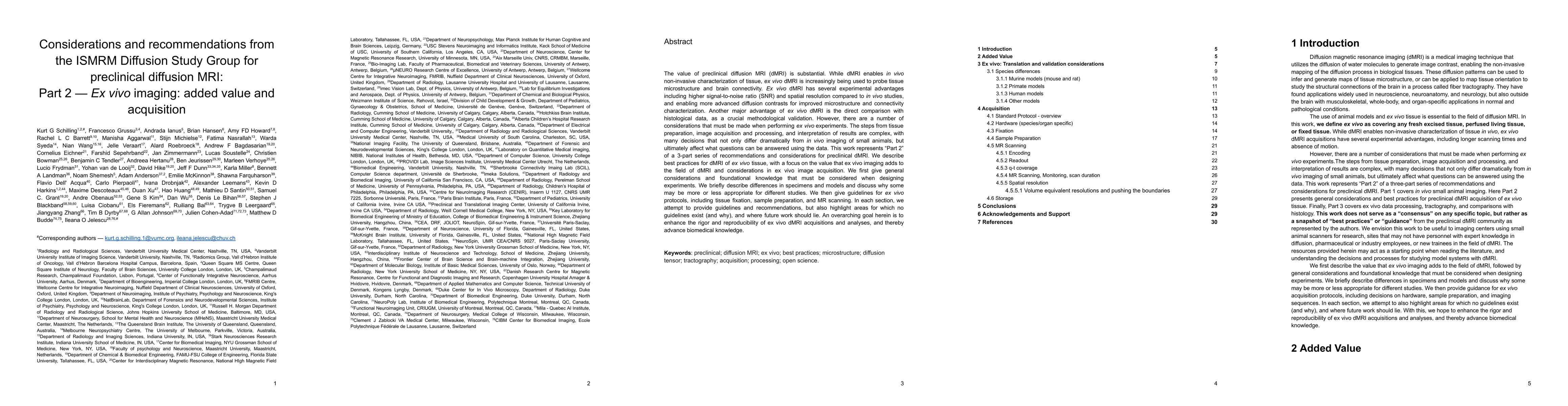
The value of preclinical diffusion MRI (dMRI) is substantial. While dMRI enables in vivo non-invasive characterization of tissue, ex vivo dMRI is increasingly being used to probe tissue microstructure and brain connectivity. Ex vivo dMRI has several experimental advantages including higher signal-to-noise ratio and spatial resolution compared to in vivo studies, and more advanced diffusion contrasts for improved microstructure and connectivity characterization. Another major advantage is direct comparison with histological data as a crucial methodological validation. However, there are a number of considerations that must be made when performing ex vivo experiments. The steps from tissue preparation, image acquisition and processing, and interpretation of results are complex, with many decisions that not only differ dramatically from in vivo imaging, but ultimately affect what questions can be answered using the data. This work represents 'Part 2' of a series of recommendations and considerations for preclinical dMRI, where we focus on best practices for dMRI of ex vivo tissue. We first describe the value that ex vivo imaging adds to the field of dMRI, followed by general considerations and foundational knowledge that must be considered when designing experiments. We then give guidelines for ex vivo protocols, including tissue preparation, imaging sequences and data processing including pre-processing, model-fitting, and tractography. Finally, we provide an online resource which lists publicly available ex vivo dMRI datasets and dedicated software packages. In each section, we attempt to provide guidelines and recommendations, but also highlight areas for which no guidelines exist, and where future work should lie. An overarching goal herein is to enhance the rigor and reproducibility of ex vivo dMRI acquisitions and analyses, and thereby advance biomedical knowledge.
This graph shows how this paper is connected to other research. The current paper is shown in dark gray, papers that cite it are in green, and papers it references are in blue. You can:
• Hover over any node to see the paper title
• Click a node to view that paper's details
• Drag nodes to rearrange the graph
• Use the zoom controls (bottom right) to zoom in/out
Title | Authors | Year | Actions |
---|---|---|---|
Non-invasive MRI of Blood-Cerebrospinal Fluid-Barrier Function: a Functional Biomarker of Early Alzheimer’s Disease Pathology
|
C. Perera, Renata Cruz, N. Shemesh, Tânia Carvalho, David L. Thomas, Jack Wells, A. Ianuş
|
2024
|
View Paper |
Quantitative diffusion imaging and genotype‐by‐sex interactions in a rat model of Alexander disease
|
N. A. Stowe, Ajay P Singh, Brian R. Barnett, Sue Y. Yi, Paloma C Frautschi, Albee Messing, Tracy L. Hagemann, John-Paul J. Yu
|
2023
|
View Paper |
Volume electron microscopy in injured rat brain validates white matter microstructure metrics from diffusion MRI
|
Ricardo Coronado-Leija, A. Abdollahzadeh, Hong-Hsi Lee, S. Coelho, B. Ades-aron, Ying Liao, R. Salo, Jussi Tohka, Alejandra Sierra, Dmitry S. Novikov, E. Fieremans
|
2023
|
View Paper |
versaFlow: a versatile pipeline for resolution adapted diffusion MRI processing and its application to studying the variability of the PRIME-DE database
|
A. Caron, A. Shmuel, Ziqi Hao, M. Descoteaux
|
2023
|
View Paper |
Mapping acute neuroinflammation in vivo with diffusion-MRI in rats given a systemic lipopolysaccharide challenge
|
Eugene Kim, I. C. Figueiredo, C. Simmons, K. Randall, L. Gonzalez, Tobias C. Wood, B. Ranieri, Paula Sureda-Gibert, O. Howes, C. Pariante, O. Pasternak, F. Dell’Acqua, F. Turkheimer, D. Cash
|
2023
|
View Paper |
Cortical cerebrovascular and metabolic perturbations in the 5xFAD mouse model of Alzheimer’s disease
|
Amandine Jullienne, J. Szu, Ryan Quan, Michelle V Trinh, Tannoz Norouzi, B. Noarbe, Amanda A. Bedwell, Kierra Eldridge, Scott C. Persohn, P. Territo, A. Obenaus
|
2023
|
View Paper |
Differentiation of white matter histopathology using b-tensor encoding and machine learning
|
Ricardo Rios-Carrillo, Alonso Ramírez-Manzanares, H. Luna-Munguía, M. Regalado, L. Concha
|
2023
|
View Paper |
Title | Authors | Year | Actions |
---|---|---|---|
Considerations and recommendations from the ISMRM Diffusion Study Group for preclinical diffusion MRI: Part 3-Ex vivo imaging: Data processing, comparisons with microscopy, and tractography.
|
K. Schilling, Amy F D Howard, Francesco Grussu, A. Ianuş, Brian Hansen, Rachel L. C. Barrett, M. Aggarwal, S. Michielse, Fatima Nasrallah, W. Syeda, Nian Wang, J. Veraart, A. Roebroeck, Andrew F Bagdasarian, C. Eichner, F. Sepehrband, Jan Zimmermann, L. Soustelle, Christien Bowman, Benjamin C. Tendler, Andreea Hertanu, B. Jeurissen, M. Verhoye, L. Frydman, Y. van de Looij, David Hike, Jeff F. Dunn, Karla L. Miller, Bennett A Landman, N. Shemesh, Adam Anderson, Emilie McKinnon, Shawna Farquharson, F. Dell'Acqua, C. Pierpaoli, I. Drobnjak, A. Leemans, K. Harkins, M. Descoteaux, Duan Xu, Hao Huang, Mathieu D. Santin, Samuel C. Grant, Andre Obenaus, Gene S Kim, Dan Wu, D. Le Bihan, S. Blackband, Luisa Ciobanu, E. Fieremans, Ruiliang Bai, T. Leergaard, Jiangyang Zhang, T. Dyrby, G. A. Johnson, Julien Cohen-Adad, M. Budde, I. Jelescu
|
2025
|
View Paper |
Considerations and recommendations from the ISMRM diffusion study group for preclinical diffusion MRI: Part 1: In vivo small-animal imaging.
|
Ileana O. Jelescu, Francesco Grussu, A. Ianuş, B. Hansen, M. Aggarwal, S. Michielse, F. Nasrallah, W. Syeda, Nian Wang, J. Veraart, A. Roebroeck, Andrew F Bagdaserian, C. Eichner, F. Sepehrband, Jan Zimmermann, B. Jeurissen, L. Frydman, Y. Looij, David Hike, J. Dunn, K. Miller, B. Landman, N. Shemesh, A. Anderson, Emilie T. McKinnon, Shawna Farquharson, F. Dell’Acqua, C. Pierpaoli, I. Drobnjak, A. Leemans, K. Harkins, M. Descoteaux, Duan Xu, M. Santin, S. Grant, A. Obenaus, Gene S Kim, Dan Wu, D. Bihan, S. Blackband, L. Ciobanu, E. Fieremans, Ruiliang Bai, T. Leergaard, Jiangyang Zhang, T. Dyrby, G. A. Johnson, J. Cohen-Adad, M. Budde, K. Schilling
|
2022
|
View Paper |
The BigMac dataset: an open resource combining multi-contrast MRI and microscopy in the macaque brain
|
A. Howard, I. Huszar, Adele Smart, M. Cottaar, G. Daubney, Taylor Hanayik, A. Khrapitchev, R. Mars, J. Mollink, Connor Scott, N. Sibson, J. Sallet, S. Jbabdi, K. Miller
|
2022
|
View Paper |
A connectomics-based taxonomy of mammals
|
Laura E. Suárez, Y. Yovel, M. P. van den Heuvel, O. Sporns, Y. Assaf, Guillaume Lajoie, B. Mišić
|
2022
|
View Paper |
Insights from the IronTract challenge: Optimal methods for mapping brain pathways from multi-shell diffusion MRI
|
Chiara Maffei, G. Girard, K. Schilling, B. Aydogan, N. Aduluru, A. Zhylka, Ye Wu, M. Mancini, A. Hamamci, Alessia Sarica, Achille Telliac, S. Baete, D. Karimi, Frank Yeh, M. Yıldız, A. Gholipour, Yann Bihan-Poudec, B. Hiba, A. Quattrone, A. Quattrone, Tommy Boshkovski, N. Stikov, P. Yap, A. de Luca, J. Pluim, A. Leemans, V. Prabhakaran, B. Bendlin, A. Alexander, B. Landman, Erick Jorge Canales-Rodríguez, M. Barakovic, Jonathan Rafael-Patiño, Thomas Yu, Gaëtan Rensonnet, S. Schiavi, Alessandro Daducci, M. Pizzolato, E. Fischi-Gómez, J. Thiran, G. Dai, Giorgia Grisot, Nikola Lazovski, Santi Puch, M. Ramos, P. Rodrigues, Vesna Prchkovska, Robert J. Jones, Julia F. Lehman, S. Haber, A. Yendiki
|
2021
|
View Paper |
Tissue Optimisation Strategies for High Quality Ex Vivo Diffusion Imaging
|
Rachel L. C. Barrett, D. Cash, C. Simmons, Eugene Kim, Tobias C. Wood, Richard Stones, A. Vernon, M. Catani, F. Dell’Acqua
|
2021
|
View Paper |
Microstructural Tissue Changes in a Rat Model of Mild Traumatic Brain Injury
|
K. Chary, Omar Narvaez, R. Salo, Isabel San Martín Molina, Jussi Tohka, M. Aggarwal, O. Gröhn, A. Sierra
|
2021
|
View Paper |
High-resolution mapping and digital atlas of subcortical regions in the macaque monkey based on matched MAP-MRI and histology
|
K. Saleem, A. Avram, D. Glen, C. Yen, F. Ye, M. Komlosh, P. Basser
|
2021
|
View Paper |
A time‐course study of actively stained mouse brains: Diffusion tensor imaging parameters and connectomic stability over 1 year
|
Jaclyn Xiao, Kathryn J. Hornburg, G. Cofer, James J. Cook, Forrest Pratson, Y. Qi, G. A. Johnson
|
2021
|
View Paper |
A method to remove the influence of fixative concentration on postmortem T2 maps using a kinetic tensor model
|
Benjamin C. Tendler, Feng Qi, S. Foxley, M. Pallebage-Gamarallage, R. Menke, O. Ansorge, S. Hurley, K. Miller
|
2021
|
View Paper |
Diffusion time dependence, power-law scaling, and exchange in gray matter
|
J. Olesen, L. Østergaard, N. Shemesh, S. Jespersen
|
2021
|
View Paper |
Neurite Exchange Imaging (NEXI): A minimal model of diffusion in gray matter with inter-compartment water exchange
|
Ileana O. Jelescu, Alexandre de Skowronski, F. Geffroy, M. Palombo, D. Novikov
|
2021
|
View Paper |
A multicontrast MR atlas of the Wistar rat brain
|
G. Johnson, Rick Laoprasert, R. J. Anderson, G. Cofer, James J. Cook, Forrest Pratson, L. White
|
2021
|
View Paper |
The Digital Brain Bank, an open access platform for post-mortem imaging datasets
|
Benjamin C. Tendler, Taylor Hanayik, O. Ansorge, Sarah Bangerter-Christensen, G. Berns, M. Bertelsen, K. Bryant, S. Foxley, M. P. van den Heuvel, A. Howard, I. Huszar, A. Khrapitchev, A. Leonte, P. Manger, R. Menke, J. Mollink, Duncan Mortimer, M. Pallebage-Gamarallage, Léa Roumazeilles, J. Sallet, L. H. Scholtens, Connor Scott, Adele Smart, M. R. Turner, Chaoyue Wang, S. Jbabdi, R. Mars, K. Miller
|
2021
|
View Paper |
Diffusion MRI and anatomic tracing in the same brain reveal common failure modes of tractography
|
Giorgia Grisot, S. Haber, A. Yendiki
|
2021
|
View Paper |
The squirrel monkey model in clinical neuroscience
|
Julie Royo, S. Forkel, P. Pouget, M. T. Schotten
|
2021
|
View Paper |
A high-resolution interactive atlas of the human brainstem using magnetic resonance imaging
|
Syed M. Adil, E. Calabrese, Lefko T. Charalambous, James J. Cook, S. Rahimpour, A. Atik, G. Cofer, Beth A. Parente, G. Johnson, S. Lad, L. White
|
2021
|
View Paper |
Tractography dissection variability: What happens when 42 groups dissect 14 white matter bundles on the same dataset?
|
K. Schilling, F. Rheault, L. Petit, Colin B. Hansen, V. Nath, F. Yeh, G. Girard, M. Barakovic, Jonathan Rafael-Patino, Thomas Yu, E. Fischi-Gómez, M. Pizzolato, Mario Ocampo-Pineda, S. Schiavi, Erick Jorge Canales-Rodríguez, Alessandro Daducci, C. Granziera, G. Innocenti, J. Thiran, L. Mancini, S. Wastling, S. Cocozza, M. Petracca, G. Pontillo, M. Mancini, S. Vos, V. Vakharia, J. Duncan, Helena Melero, Lidia Manzanedo, Emilio Sanz-Morales, Á. Peña-Melián, F. Calamante, A. Attyé, R. Cabeen, L. Korobova, A. Toga, A. Vijayakumari, D. Parker, R. Verma, A. Radwan, S. Sunaert, L. Emsell, A. de Luca, A. Leemans, C. Bajada, H. Haroon, H. Azadbakht, Maxime Chamberland, Sila Genc, C. Tax, P. Yeh, R. Srikanchana, C. McKnight, J. Yang, Jian Chen, Claire E. Kelly, Chun-Hung Yeh, Jérôme Cochereau, J. Maller, T. Welton, F. Almairac, K. Seunarine, C. Clark, Fan Zhang, N. Makris, A. Golby, Y. Rathi, L. O’Donnell, Yihao Xia, D. Aydogan, Yonggang Shi, F. G. Fernandes, M. Raemaekers, S. Warrington, S. Michielse, Alonso Ramírez-Manzanares, L. Concha, Ramón Aranda, M. Meraz, Garikoitz Lerma-Usabiaga, L. Roitman, L. Fekonja, Navona Calarco, Michael J. E. Joseph, Hajer Nakua, A. Voineskos, Philippe Karan, G. Grenier, J. Legarreta, N. Adluru, Veena A. Nair, V. Prabhakaran, A. Alexander, K. Kamagata, Y. Saito, W. Uchida, C. Andica, Abe Masahiro, Roza G. Bayrak, C. G. Gandini Wheeler-Kingshott, E. D’Angelo, Fulvia Palesi, G. Savini, N. Rolandi, P. Guevara, J. Houenou, N. López-López, J. F. Mangin, C. Poupon, C. Román, Andrea Vázquez, Chiara Maffei, M. Arantes, J. P. Andrade, S. M. Silva, Rajikha Raja, V. Calhoun, E. Caverzasi, Simone Sacco, M. Lauricella, F. Pestilli, D. Bullock, Yang Zhan, E. Brignoni-Pérez, C. Lebel, Jess E. Reynolds, I. Nestrašil, R. Labounek, C. Lenglet, Amy Paulson, Štefánia Aulická, Sarah R. Heilbronner, K. Heuer, A. Anderson, B. Landman, M. Descoteaux
|
2020
|
View Paper |
Brain connections derived from diffusion MRI tractography can be highly anatomically accurate—if we know where white matter pathways start, where they end, and where they do not go
|
K. Schilling, L. Petit, F. Rheault, Samuel W. Remedios, C. Pierpaoli, A. Anderson, B. Landman, M. Descoteaux
|
2020
|
View Paper |
Challenges for biophysical modeling of microstructure
|
I. Jelescu, M. Palombo, F. Bagnato, K. Schilling
|
2020
|
View Paper |
Mesoscale diffusion magnetic resonance imaging of the ex vivo human hippocampus
|
M. Ly, L. Foley, Ashwinee Manivannan, T. K. Hitchens, R. Richardson, M. Modo
|
2020
|
View Paper |
Genetic and microstructural differences in the cortical plate of gyri and sulci during gyrification in fetal sheep.
|
Sebastian Quezada, Y. van de Looij, N. Hale, Shreya Rana, S. Sizonenko, Courtney P. Gilchrist, M. Castillo-Melendez, M. Tolcos, D. Walker
|
2020
|
View Paper |
On the Origins of Diffusion MRI Signal Changes in Stroke
|
S. Blackband, J. Flint, B. Hansen, T. Shepherd, C. Lee, W. Streit, J. Forder
|
2020
|
View Paper |
Conservation of brain connectivity and wiring across the mammalian class
|
Y. Assaf, Arieli Bouznach, Omri Zomet, A. Marom, Y. Yovel
|
2020
|
View Paper |
Visualization of live, mammalian neurons during Kainate-infusion using magnetic resonance microscopy
|
J. Flint, K. Menon, B. Hansen, J. Forder, S. Blackband
|
2020
|
View Paper |
Axon morphology is modulated by the local environment and impacts the noninvasive investigation of its structure–function relationship
|
M. Andersson, H. Kjer, Jonathan Rafael-Patino, A. Pacureanu, B. Pakkenberg, J. Thiran, M. Ptito, M. Bech, A. Dahl, Vedrana Andersen Dahl, T. Dyrby
|
2020
|
View Paper |
Virtual mouse brain histology from multi-contrast MRI via deep learning
|
Zifei Liang, C. Lee, Tanzil Mahmud Arefin, Zijun Dong, P. Walczak, S. Shi, F. Knoll, Y. Ge, L. Ying, Jiangyang Zhang
|
2020
|
View Paper |
Cytoarchitecture of the mouse brain by high resolution diffusion magnetic resonance imaging
|
Nian Wang, Leonard E. White, Y. Qi, G. Cofer, G. A. Johnson
|
2020
|
View Paper |
Ex vivo diffusion-weighted MRI tractography of the Göttingen minipig limbic system
|
Johannes Bech, Dariusz Orlowski, A. Glud, T. Dyrby, J. Sørensen, C. Bjarkam
|
2020
|
View Paper |
Longitudinal assessment of tissue properties and cardiac diffusion metrics of the ex vivo porcine heart at 7 T: Impact of continuous tissue fixation using formalin
|
D. Lohr, M. Terekhov, F. Veit, L. Schreiber
|
2020
|
View Paper |
Insight into the fundamental trade-offs of diffusion MRI from polarization-sensitive optical coherence tomography in ex vivo human brain
|
Robert J. Jones, Giorgia Grisot, J. Augustinack, C. Magnain, D. Boas, B. Fischl, Hui Wang, A. Yendiki
|
2020
|
View Paper |
Diffusion‐time dependence of diffusional kurtosis in the mouse brain
|
M. Aggarwal, Matthew D. Smith, P. Calabresi
|
2020
|
View Paper |
Adaptive phase correction of diffusion-weighted images
|
M. Pizzolato, G. Gilbert, J. Thiran, M. Descoteaux, R. Deriche
|
2020
|
View Paper |
Magnetic Resonance Microscopy at Cellular Resolution and Localised Spectroscopy of Medicago truncatula at 22.3 Tesla
|
R. van Schadewijk, J. R. Krug, Defeng Shen, Karthick B. S. Sankar Gupta, F. Vergeldt, T. Bisseling, A. Webb, H. Van As, A. Velders, H. D. de Groot, A. Alia
|
2020
|
View Paper |
Modeling an equivalent b‐value in diffusion‐weighted steady‐state free precession
|
Benjamin C. Tendler, S. Foxley, M. Cottaar, S. Jbabdi, K. Miller
|
2020
|
View Paper |
Validation of structural brain connectivity networks: The impact of scanning parameters
|
Karen Sandø Ambrosen, S. Eskildsen, M. Hinne, K. Krug, H. Lundell, Mikkel N. Schmidt, M. Gerven, Morten Mørup, T. Dyrby
|
2020
|
View Paper |
A resource for detailed 3D mapping of white matter pathways in the marmoset brain
|
Cirong Liu, F. Ye, J. Newman, Diego Szczupak, Xiaoguang Tian, C. Yen, P. Majka, D. Glen, M. Rosa, D. Leopold, Afonso C. Silva
|
2019
|
View Paper |
Influence of water based embedding media composition on the relaxation properties of fixed tissue.
|
I. Vučković, Tarek Nayfeh, P. Mishra, Sigapriya Periyanan, C. Sussman, T. Kline, S. Macura
|
2019
|
View Paper |
Ultra-high resolution and multi-shell diffusion MRI of intact ex vivo human brains using kT-dSTEAM at 9.4T
|
Francisco J. Fritz, S. Sengupta, R. Harms, D. Tse, B. Poser, A. Roebroeck
|
2019
|
View Paper |
In vivo Diffusion Tensor Magnetic Resonance Tractography of the Sheep Brain: An Atlas of the Ovine White Matter Fiber Bundles
|
V. Pieri, M. Trovatelli, M. Cadioli, D. Zani, S. Brizzola, G. Ravasio, F. Acocella, M. Di Giancamillo, L. Malfassi, M. Dolera, M. Riva, L. Bello, A. Falini, A. Castellano
|
2019
|
View Paper |
Waxholm Space atlas of the rat brain auditory system: Three-dimensional delineations based on structural and diffusion tensor magnetic resonance imaging
|
K. Osen, Jala Imad, Arvind E. Wennberg, Eszter A. Papp, T. Leergaard
|
2019
|
View Paper |
Perfusion fixation in brain banking: a systematic review
|
W. C. McFadden, Hadley Walsh, Felix Richter, Céline Soudant, C. Bryce, P. Hof, M. Fowkes, J. Crary, Andrew T. McKenzie
|
2019
|
View Paper |
Correlation tensor magnetic resonance imaging
|
R. Henriques, S. Jespersen, N. Shemesh
|
2019
|
View Paper |
Multidimensional diffusion MRI with spectrally modulated gradients reveals unprecedented microstructural detail
|
H. Lundell, M. Nilsson, T. Dyrby, T. Dyrby, G. Parker, P. Cristinacce, F. Zhou, D. Topgaard, S. Lasič
|
2019
|
View Paper |
Commentary: The Nomenclature of Human White Matter Association Pathways: Proposal for a Systematic Taxonomic Anatomical Classification
|
S. Panesar, J. Fernandez-Miranda
|
2019
|
View Paper |
The dot-compartment revealed? Diffusion MRI with ultra-strong gradients and spherical tensor encoding in the living human brain
|
C. Tax, Filip Szczepankiewicz, M. Nilsson, Derek K. Jones
|
2019
|
View Paper |
Uncovering the inferior fronto-occipital fascicle and its topological organization in non-human primates: the missing connection for language evolution
|
S. Sarubbo, L. Petit, A. De Benedictis, F. Chioffi, M. Ptito, T. Dyrby
|
2019
|
View Paper |
Post mortem high resolution diffusion MRI for large specimen imaging at 11.7 T with 3D segmented echo-planar imaging
|
Sophie B. Sébille, Anne-Sophie Rolland, M. Welter, É. Bardinet, M. Santin
|
2019
|
View Paper |
Comparison between diffusion MRI tractography and histological tract-tracing of cortico-cortical structural connectivity in the ferret brain
|
C. Delettre, A. Messé, L. Dell, O. Foubet, K. Heuer, B. Larrat, S. Mériaux, J. F. Mangin, I. Reillo, C. de Juan Romero, V. Borrell, R. Toro, C. Hilgetag
|
2019
|
View Paper |
The Nomenclature of Human White Matter Association Pathways: Proposal for a Systematic Taxonomic Anatomical Classification
|
E. Mandonnet, S. Sarubbo, L. Petit
|
2018
|
View Paper |
Physical and numerical phantoms for the validation of brain microstructural MRI: A cookbook
|
E. Fieremans, Hong-Hsi Lee
|
2018
|
View Paper |
Validation strategies for the interpretation of microstructure imaging using diffusion MRI
|
T. Dyrby, G. Innocenti, M. Bech, H. Lundell
|
2018
|
View Paper |
Microstructural imaging of the human brain with a ‘super-scanner’: 10 key advantages of ultra-strong gradients for diffusion MRI
|
Derek K. Jones, Derek K. Jones, Daniel C. Alexander, Daniel C. Alexander, Richard Bowtell, Mara Cercignani, F. Dell'Acqua, Damien J. McHugh, Damien J. McHugh, Karla L. Miller, Marco Palombo, Geoffrey J. M. Parker, Geoffrey J. M. Parker, S. Rudrapatna, C. Tax
|
2018
|
View Paper |
Evidence of intraneuronal Aβ accumulation preceding tau pathology in the entorhinal cortex
|
L. Welikovitch, S. Do Carmo, Z. Maglóczky, P. Szocsics, J. Loke, T. Freund, A. C. Cuello
|
2018
|
View Paper |
Effect of Fluorinert on the Histological Properties of Formalin-Fixed Human Brain Tissue
|
J. E. Iglesias, Shauna Crampsie, C. Strand, M. Tachrount, D. Thomas, J. Holton
|
2018
|
View Paper |
Detection and Distinction of Mild Brain Injury Effects in a Ferret Model Using Diffusion Tensor MRI (DTI) and DTI-Driven Tensor-Based Morphometry (D-TBM)
|
Elizabeth B. Hutchinson, Elizabeth B. Hutchinson, S. Schwerin, S. Schwerin, Kryslaine L. Radomski, Kryslaine L. Radomski, N. Sadeghi, M. Komlosh, M. Komlosh, M. Irfanoglu, S. Juliano, C. Pierpaoli
|
2018
|
View Paper |
Micro and Nano Scale NMR
|
J. Anders, J. Korvink
|
2018
|
View Paper |
Ex vivo diffusion MRI of the human brain: Technical challenges and recent advances
|
A. Roebroeck, K. Miller, M. Aggarwal
|
2018
|
View Paper |
A digital 3D atlas of the marmoset brain based on multi-modal MRI
|
Cirong Liu, F. Ye, C. Yen, J. Newman, D. Glen, D. Leopold, Afonso C. Silva
|
2018
|
View Paper |
Quantitative Ex Vivo MRI Changes due to Progressive Formalin Fixation in Whole Human Brain Specimens: Longitudinal Characterization of Diffusion, Relaxometry, and Myelin Water Fraction Measurements at 3T
|
A. Shatil, M. Uddin, K. Matsuda, C. Figley
|
2018
|
View Paper |
Effects of concentration and vendor specific composition of formalin on postmortem MRI of the human brain
|
C. Birkl, M. Soellradl, A. M. Toeglhofer, S. Krassnig, M. Leoni, L. Pirpamer, Thomas Vorauer, H. Krenn, J. Haybaeck, F. Fazekas, S. Ropele, C. Langkammer
|
2018
|
View Paper |
Histological validation of diffusion MRI fiber orientation distributions and dispersion
|
K. Schilling, Vaibhav A. Janve, Yurui Gao, I. Stepniewska, B. Landman, A. Anderson
|
2018
|
View Paper |
Accurate estimation of microscopic diffusion anisotropy and its time dependence in the mouse brain
|
A. Ianuş, S. Jespersen, Teresa Serradas Duarte, D. Alexander, I. Drobnjak, N. Shemesh
|
2017
|
View Paper |
Precision and accuracy of diffusion kurtosis estimation and the influence of b‐value selection
|
A. Chuhutin, B. Hansen, S. Jespersen
|
2017
|
View Paper |
Can increased spatial resolution solve the crossing fiber problem for diffusion MRI?
|
K. Schilling, Yurui Gao, Vaibhav A. Janve, I. Stepniewska, B. Landman, A. Anderson
|
2017
|
View Paper |
Microscopic Magnetic Resonance Imaging: A Practical Perspective
|
L. Ciobanu
|
2017
|
View Paper |
Neurite dispersion: a new marker of multiple sclerosis spinal cord pathology?
|
F. Grussu, T. Schneider, C. Tur, Richard L Yates, M. Tachrount, A. Ianuş, M. Yiannakas, J. Newcombe, Hui Zhang, D. Alexander, G. DeLuca, C. G. Gandini Wheeler-Kingshott
|
2017
|
View Paper |
Population-averaged macaque brain atlas with high-resolution ex vivo DTI integrated into in vivo space
|
Lei Feng, T. Jeon, Q. Yu, M. Ouyang, Qinmu Peng, V. Mishra, M. Pletikos, N. Šestan, M. Miller, S. Mori, Steven Hsiao, Shuwei Liu, Hao Huang
|
2017
|
View Paper |
Exploring sex differences in the adult zebra finch brain: In vivo diffusion tensor imaging and ex vivo super-resolution track density imaging
|
J. Hamaide, G. Groof, G. Steenkiste, B. Jeurissen, J. Audekerke, M. Naeyaert, L. V. Ruijssevelt, C. Cornil, Jan Sijbers, M. Verhoye, A. Linden
|
2017
|
View Paper |
Multidimensional diffusion MRI.
|
Daniel Topgaard
|
2017
|
View Paper |
The importance of correcting for signal drift in diffusion MRI
|
S. Vos, C. Tax, P. Luijten, S. Ourselin, A. Leemans, M. Froeling
|
2017
|
View Paper |
Gradient and shim technologies for ultra high field MRI
|
S. Winkler, F. Schmitt, H. Landes, Josh DeBever, T. Wade, A. Alejski, B. Rutt
|
2016
|
View Paper |
Variations in post-perfusion immersion fixation and storage alter MRI measurements of mouse brain morphometry
|
A. de Guzman, M. Wong, J. Gleave, B. Nieman
|
2016
|
View Paper |
Quantitative MRI and DTI Abnormalities During the Acute Period Following CCI in the Ferret
|
E. Hutchinson, S. Schwerin, Kryslaine L. Radomski, M. Irfanoglu, S. Juliano, C. Pierpaoli
|
2016
|
View Paper |
In vivo quantification of demyelination and recovery using compartment-specific diffusion MRI metrics validated by electron microscopy
|
Ileana O. Jelescu, M. Zurek, Kerryanne V. Winters, J. Veraart, Anjali Rajaratnam, Nathanael S. Kim, J. Babb, T. Shepherd, D. Novikov, S. Kim, E. Fieremans
|
2016
|
View Paper |
Regional T1 relaxation time constants in Ex vivo human brain: Longitudinal effects of formalin exposure
|
Mekala R. Raman, Y. Shu, T. Lesnick, C. Jack, K. Kantarci
|
2016
|
View Paper |
Towards higher sensitivity and stability of axon diameter estimation with diffusion‐weighted MRI
|
F. Sepehrband, D. Alexander, N. Kurniawan, D. Reutens, Zhengyi Yang
|
2016
|
View Paper |
Evaluation of diffusion kurtosis imaging in ex vivo hypomyelinated mouse brains
|
Nathaniel D. Kelm, Kathryn L. West, Robert P. Carson, D. Gochberg, K. Ess, M. Does
|
2016
|
View Paper |
A Microperfusion and In-Bore Oxygenator System Designed for Magnetic Resonance Microscopy Studies on Living Tissue Explants
|
J. Flint, K. Menon, B. Hansen, J. Forder, S. Blackband
|
2015
|
View Paper |
Extracellular space preservation aids the connectomic analysis of neural circuits
|
Marta Pallotto, P. V. Watkins, B. Fubara, J. Singer, K. Briggman
|
2015
|
View Paper |
The extreme capsule fiber complex in humans and macaque monkeys: a comparative diffusion MRI tractography study
|
R. Mars, S. Foxley, L. Verhagen, S. Jbabdi, J. Sallet, M. Noonan, Franz-Xaver Neubert, J. Andersson, P. Croxson, Robin I. M. Dunbar, A. Khrapitchev, N. Sibson, K. Miller, M. Rushworth
|
2015
|
View Paper |
Ultrastructural analysis of adult mouse neocortex comparing aldehyde perfusion with cryo fixation
|
N. Korogod, C. Petersen, G. Knott
|
2015
|
View Paper |
Postmortem diffusion MRI of the human brainstem and thalamus for deep brain stimulator electrode localization
|
E. Calabrese, Patrick Hickey, C. Hulette, Jingxian Zhang, Beth A. Parente, S. Lad, G. Johnson
|
2015
|
View Paper |
Histological validation of high-resolution DTI in human post mortem tissue
|
A. Seehaus, A. Roebroeck, M. Bastiani, L. Fonseca, H. Bratzke, N. Lori, A. Vilanova, R. Goebel, R. Galuske
|
2015
|
View Paper |
Investigation of the subcellular architecture of L7 neurons of Aplysia californica using magnetic resonance microscopy (MRM) at 7.8 microns
|
C. Lee, J. Flint, B. Hansen, S. Blackband
|
2015
|
View Paper |
A Diffusion MRI Tractography Connectome of the Mouse Brain and Comparison with Neuronal Tracer Data
|
E. Calabrese, A. Badea, G. Cofer, Y. Qi, G. Johnson
|
2015
|
View Paper |
Neurite orientation dispersion and density imaging of the healthy cervical spinal cord in vivo
|
Francesco Grussu, T. Schneider, Hui Zhang, D. Alexander, C. Wheeler-Kingshott
|
2015
|
View Paper |
Assessment of murine brain tissue shrinkage caused by different histological fixatives using magnetic resonance and computed tomography imaging.
|
H. Wehrl, I. Bezrukov, S. Wiehr, M. Lehnhoff, K. Fuchs, J. Mannheim, L. Quintanilla‐Martinez, U. Kohlhofer, M. Kneilling, B. Pichler, A. Sauter
|
2015
|
View Paper |
Visualization of synaptic domains in the Drosophila brain by magnetic resonance microscopy at 10 micron isotropic resolution
|
C. Lee, S. Blackband, P. Fernandez-Funez
|
2015
|
View Paper |
Waxholm Space atlas of the rat brain hippocampal region: Three-dimensional delineations based on magnetic resonance and diffusion tensor imaging
|
Lisa J. Kjonigsen, S. Lillehaug, J. Bjaalie, M. Witter, T. Leergaard
|
2015
|
View Paper |
Probing region-specific microstructure of human cortical areas using high angular and spatial resolution diffusion MRI
|
M. Aggarwal, David W. Nauen, J. Troncoso, S. Mori
|
2015
|
View Paper |
Mapping mean axon diameter and axonal volume fraction by MRI using temporal diffusion spectroscopy
|
Junzhong Xu, Hua Li, K. Harkins, Xiaoyu Jiang, Jingping Xie, Hakmook Kang, M. Does, J. Gore
|
2014
|
View Paper |
Interpolation of diffusion weighted imaging datasets
|
T. Dyrby, H. Lundell, M. Burke, N. L. Reislev, O. Paulson, M. Ptito, H. Siebner
|
2014
|
View Paper |
Improving diffusion-weighted imaging of post-mortem human brains: SSFP at 7 T
|
S. Foxley, S. Jbabdi, S. Clare, W. Lam, O. Ansorge, G. Douaud, K. Miller
|
2014
|
View Paper |
Anatomical accuracy of brain connections derived from diffusion MRI tractography is inherently limited
|
Cibu Thomas, Cibu Thomas, F. Ye, M. Irfanoglu, Pooja Modi, K. Saleem, D. Leopold, C. Pierpaoli
|
2014
|
View Paper |
Oscillating gradient diffusion MRI reveals unique microstructural information in normal and hypoxia‐ischemia injured mouse brains
|
Dan Wu, L. Martin, F. Northington, Jiangyang Zhang
|
2014
|
View Paper |
Cross-validation of serial optical coherence scanning and diffusion tensor imaging: A study on neural fiber maps in human medulla oblongata
|
Hui Wang, Junfeng Zhu, M. Reuter, L. Vinke, A. Yendiki, D. Boas, B. Fischl, T. Akkin
|
2014
|
View Paper |
Apparent exchange rate imaging in anisotropic systems
|
C. Sønderby, H. Lundell, L. V. Søgaard, T. Dyrby
|
2014
|
View Paper |
Waxholm Space atlas of the Sprague Dawley rat brain
|
Eszter A. Papp, T. Leergaard, E. Calabrese, G. Johnson, J. Bjaalie
|
2014
|
View Paper |
Relationship between the diffusion time and the diffusion MRI signal observed at 17.2 tesla in the healthy rat brain cortex
|
N. Pyatigorskaya, D. Bihan, O. Reynaud, L. Ciobanu
|
2014
|
View Paper |
Effects of hypotonic stress and ouabain on the apparent diffusion coefficient of water at cellular and tissue levels in Aplysia
|
Ileana O. Jelescu, L. Ciobanu, F. Geffroy, P. Marquet, D. Le Bihan
|
2014
|
View Paper |
A segmentation protocol and MRI atlas of the C57BL/6J mouse neocortex
|
Jeremy F. P. Ullmann, Charles Watson, A. Janke, N. Kurniawan, D. Reutens
|
2013
|
View Paper |
Feasibility of creating a high-resolution 3D diffusion tensor imaging based atlas of the human brainstem: A case study at 11.7T
|
M. Aggarwal, Jiangyang Zhang, O. Pletnikova, B. Crain, J. Troncoso, S. Mori
|
2013
|
View Paper |
Oscillating and pulsed gradient diffusion magnetic resonance microscopy over an extended b‐value range: Implications for the characterization of tissue microstructure
|
S. Portnoy, J. Flint, S. Blackband, G. J. Stanisz, G. J. Stanisz
|
2013
|
View Paper |
Diffusion Tensor Imaging of White Matter Tracts in the Dog Brain
|
O. Jacqmot, B. Van Thielen, Y. Fierens, Martha Hammond, I. Willekens, P. Schuerbeek, F. Verhelle, P. Goossens, Filip De Ridder, J. Clarys, A. Vanbinst, J. de Mey
|
2013
|
View Paper |
Quantification of anisotropy and fiber orientation in human brain histological sections
|
M. Budde, J. Annese
|
2012
|
View Paper |
Examining brain microstructure using structure tensor analysis of histological sections
|
M. Budde, J. Frank
|
2012
|
View Paper |
Contrast and stability of the axon diameter index from microstructure imaging with diffusion MRI
|
T. Dyrby, Lise V S⊘gaard, M. Hall, M. Ptito, D. Alexander
|
2012
|
View Paper |
A multidimensional magnetic resonance histology atlas of the Wistar rat brain
|
G. Johnson, E. Calabrese, A. Badea, G. Paxinos, G. Paxinos, Charles Watson, Charles Watson, Charles Watson
|
2012
|
View Paper |
High-field diffusion tensor imaging characterization of cerebral white matter injury in lipopolysaccharide-exposed fetal sheep
|
Y. Looij, G. Lodygensky, J. Dean, F. Lazeyras, H. Hagberg, I. Kjellmer, C. Mallard, P. Hüppi, S. Sizonenko
|
2012
|
View Paper |
Magnetic resonance microscopy of human and porcine neurons and cellular processes
|
J. Flint, B. Hansen, S. Portnoy, C. Lee, M. King, M. Fey, F. Vincent, G. Stanisz, P. Vestergaard-Poulsen, S. Blackband
|
2012
|
View Paper |
In vivo and ex vivo diffusion tensor imaging of cuprizone‐induced demyelination in the mouse corpus callosum
|
Jiangyang Zhang, M. Jones, M. McMahon, S. Mori, P. Calabresi
|
2012
|
View Paper |
Compartment models of the diffusion MR signal in brain white matter: A taxonomy and comparison
|
Eleftheria Panagiotaki, T. Schneider, B. Siow, M. Hall, M. Lythgoe, D. Alexander
|
2012
|
View Paper |
Diffusion tractography of post-mortem human brains: Optimization and comparison of spin echo and steady-state free precession techniques
|
K. Miller, J. McNab, S. Jbabdi, G. Douaud
|
2012
|
View Paper |
Comparison of In Vivo and Ex Vivo Diffusion Tensor Imaging in Rhesus Macaques at Short and Long Diffusion Times
|
S. Rane, T. Duong
|
2011
|
View Paper |
Population-averaged diffusion tensor imaging atlas of the Sprague Dawley rat brain
|
J. Veraart, T. Leergaard, Bjørnar T. Antonsen, W. Hecke, I. Blockx, B. Jeurissen, Yi Jiang, A. Linden, G. Johnson, M. Verhoye, Jan Sijbers
|
2011
|
View Paper |
Diffusion tensor microscopy in human nervous tissue with quantitative correlation based on direct histological comparison
|
B. Hansen, J. Flint, C. Lee, M. Fey, F. Vincent, M. King, P. Vestergaard-Poulsen, S. Blackband
|
2011
|
View Paper |
Diffusion imaging of whole, post-mortem human brains on a clinical MRI scanner
|
K. Miller, C. Stagg, G. Douaud, S. Jbabdi, Stephen M. Smith, Timothy Edward John Behrens, M. Jenkinson, S. Chance, M. Esiri, N. Voets, N. Jenkinson, T. Aziz, M. Turner, H. Johansen-Berg, J. McNab
|
2011
|
View Paper |
An ex vivo imaging pipeline for producing high‐quality and high‐resolution diffusion‐weighted imaging datasets
|
T. Dyrby, W. Baaré, D. Alexander, J. Jelsing, E. Garde, L. V. Søgaard
|
2011
|
View Paper |
Phase errors in Diffusion-Weighted Imaging
|
R. Bammer, S. Holdsworth, M. Aksoy, S. Skare
|
2010
|
View Paper |
Waxholm Space: An image-based reference for coordinating mouse brain research
|
G. Johnson, A. Badea, J. Brandenburg, G. Cofer, B. Fubara, Song Liu, J. Nissanov, J. Nissanov
|
2010
|
View Paper |
Insights into Diffusion Tensor Imaging from Animal Models of White Matter Pathology
|
M. Budde, Sheng-Kwei Song
|
2010
|
View Paper |
Orientationally invariant indices of axon diameter and density from diffusion MRI
|
D. Alexander, P. Cristinacce, M. Hall, E. Moore, M. Ptito, G. Parker, T. Dyrby
|
2010
|
View Paper |
Cellular-level diffusion tensor microscopy and fiber tracking in mammalian nervous tissue with direct histological correlation
|
J. Flint, B. Hansen, M. Fey, Daniel Schmidig, M. King, P. Vestergaard-Poulsen, S. Blackband
|
2010
|
View Paper |
Longitudinal characterization of brain atrophy of a Huntington's disease mouse model by automated morphological analyses of magnetic resonance images
|
Jiangyang Zhang, Qi Peng, Qing Li, N. Jahanshad, Zhipeng Hou, Mali Jiang, N. Masuda, D. Langbehn, M. Miller, S. Mori, C. Ross, W. Duan
|
2010
|
View Paper |
Neurite density from magnetic resonance diffusion measurements at ultrahigh field: Comparison with light microscopy and electron microscopy
|
S. Jespersen, C. Bjarkam, J. Nyengaard, M. Chakravarty, B. Hansen, T. Vosegaard, L. Østergaard, D. Yablonskiy, N. C. Nielsen, P. Vestergaard-Poulsen
|
2010
|
View Paper |
Quantitative characterization of tissue microstructure with temporal diffusion spectroscopy.
|
Junzhong Xu, M. Does, J. Gore
|
2009
|
View Paper |
The pig model in brain imaging and neurosurgery.
|
P. Sauleau, E. Lapouble, D. Val-Laillet, C. Malbert
|
2009
|
View Paper |
Magnetic resonance microscopy of mammalian neurons
|
J. Flint, C. Lee, B. Hansen, M. Fey, Daniel Schmidig, J. D. Bui, M. King, P. Vestergaard-Poulsen, S. Blackband
|
2009
|
View Paper |
Aldehyde fixative solutions alter the water relaxation and diffusion properties of nervous tissue
|
T. Shepherd, P. Thelwall, G. Stanisz, S. Blackband
|
2009
|
View Paper |
High resolution diffusion-weighted imaging in fixed human brain using diffusion-weighted steady state free precession
|
J. McNab, S. Jbabdi, S. Deoni, G. Douaud, Timothy Edward John Behrens, K. Miller
|
2009
|
View Paper |
Diffusion weighted magnetic resonance imaging of neuronal activity in the hippocampal slice model
|
J. Flint, B. Hansen, P. Vestergaard-Poulsen, S. Blackband
|
2009
|
View Paper |
Postmortem MRI of human brain hemispheres: T2 relaxation times during formaldehyde fixation
|
R. Dawe, David A. Bennett, J. Schneider, S. Vasireddi, K. Arfanakis
|
2009
|
View Paper |
New insights into tumor microstructure using temporal diffusion spectroscopy.
|
D. Colvin, T. Yankeelov, M. Does, Zoe Yue, C. Quarles, J. Gore
|
2008
|
View Paper |
Assessment of factors that confound MRI and neuropathological correlation of human postmortem brain tissue
|
L. Grinberg, E. Amaro, S. Teipel, Denis Dionizio dos Santos, C. Pasqualucci, R. Leite, Celia Regina Camargo, J. A. Gonçalves, Ariadne Gonçalves Sanches, Miriam Santana, R. Ferretti, W. Jacob-Filho, R. Nitrini, H. Heinsen, B. B. V. S. Group
|
2008
|
View Paper |
Axcaliber: A method for measuring axon diameter distribution from diffusion MRI
|
Y. Assaf, T. Blumenfeld-Katzir, Y. Yovel, P. Basser
|
2008
|
View Paper |
High-resolution diffusion tensor imaging and tractography of the human optic chiasm at 9.4 T
|
A. Roebroeck, R. Galuske, E. Formisano, O. Chiry, H. Bratzke, I. Ronen, Dae-Shik Kim, R. Goebel
|
2008
|
View Paper |
Validation of in vitro probabilistic tractography
|
T. Dyrby, L. V. Søgaard, G. Parker, D. Alexander, N. M. Lind, W. Baaré, A. Hay-Schmidt, N. Eriksen, B. Pakkenberg, O. Paulson, J. Jelsing
|
2007
|
View Paper |
The effects of brain tissue decomposition on diffusion tensor imaging and tractography
|
H. D'Arceuil, A. Crespigny
|
2007
|
View Paper |
An approach to high resolution diffusion tensor imaging in fixed primate brain
|
H. D'Arceuil, S. Westmoreland, A. Crespigny
|
2007
|
View Paper |
Magnetic field and tissue dependencies of human brain longitudinal 1H2O relaxation in vivo
|
W. Rooney, G. Johnson, Xin Li, E. R. Cohen, Seong-Gi Kim, K. Uğurbil, C. Springer
|
2007
|
View Paper |
Reversible and irreversible effects of chemical fixation on the NMR properties of single cells
|
A. Purea, A. Webb
|
2006
|
View Paper |
High magnetic field water and metabolite proton T1 and T2 relaxation in rat brain in vivo
|
R. D. de Graaf, P. Brown, S. McIntyre, T. Nixon, K. Behar, D. Rothman
|
2006
|
View Paper |
Effects of temperature and aldehyde fixation on tissue water diffusion properties, studied in an erythrocyte ghost tissue model
|
P. Thelwall, T. Shepherd, G. Stanisz, S. Blackband
|
2006
|
View Paper |
Translational neuroscience and magnetic-resonance microscopy
|
H. Benveniste, S. Blackband
|
2006
|
View Paper |
Fiber Pathways of the Brain
|
J. Schmahmann, D. Pandya
|
2006
|
View Paper |
Statistical diffusion tensor histology reveals regional dysmyelination effects in the shiverer mouse mutant
|
J. Tyszka, C. Readhead, Elaine L. Bearer, Elaine L. Bearer, R. Pautler, R. Pautler, R. Jacobs
|
2006
|
View Paper |
Formalin fixation alters water diffusion coefficient magnitude but not anisotropy in infarcted brain
|
Shu-wei Sun, J. Neil, Hsiao-Fang Liang, Yong Y. He, R. Schmidt, C. Hsu, Sheng-Kwei Song
|
2005
|
View Paper |
Ex vivo evaluation of ADC values within spinal cord white matter tracts.
|
E. Schwartz, Emily T. Cooper, C. Chin, Suzanne Wehrli, A. Tessler, D. Hackney
|
2005
|
View Paper |
Postmortem MR imaging of formalin-fixed human brain
|
A. Pfefferbaum, E. Sullivan, E. Adalsteinsson, T. Garrick, C. Harper
|
2004
|
View Paper |
Relative indices of water diffusion anisotropy are equivalent in live and formalin‐fixed mouse brains
|
Shu-wei Sun, J. Neil, Sheng-Kwei Song
|
2003
|
View Paper |
Oscillating gradient measurements of water diffusion in normal and globally ischemic rat brain
|
M. Does, E. Parsons, J. Gore
|
2003
|
View Paper |
The basis of anisotropic water diffusion in the nervous system – a technical review
|
Christian Beaulieu
|
2002
|
View Paper |
Human erythrocyte ghosts: Exploring the origins of multiexponential water diffusion in a model biological tissue with magnetic resonance
|
P. Thelwall, S. Grant, G. Stanisz, S. Blackband
|
2002
|
View Paper |
Morphologic phenotyping with MR microscopy: the visible mouse.
|
G. Johnson, G. Cofer, S. Gewalt, L. Hedlund
|
2002
|
View Paper |
MR microscopy of multicomponent diffusion in single neurons
|
S. Grant, D. Buckley, S. Gibbs, A. Webb, S. Blackband
|
2001
|
View Paper |
Water diffusion compartmentation and anisotropy at high b values in the human brain
|
C. Clark, D. Le Bihan
|
2000
|
View Paper |
Formaldehyde, Formalin, Paraformaldehyde And Glutaraldehyde: What They Are And What They Do
|
J. Kiernan
|
2000
|
View Paper |
Color schemes to represent the orientation of anisotropic tissues from diffusion tensor data: Application to white matter fiber tract mapping in the human brain
|
S. Pajevic, C. Pierpaoli
|
1999
|
View Paper |
A Fixation Procedure for Ultrastructural Investigation of Synaptic Connections in Resected Human Cortex
|
F. Morin, C. Crevier, G. Bouvier, J. Lacaille, C. Beaulieu
|
1997
|
View Paper |
A study of diffusion isotropy in single neurons by using NMR microscopy
|
E. Hsu, N. Aiken, S. Blackband
|
1997
|
View Paper |
Nuclear magnetic resonance microscopy of single neurons under hypotonic perturbation.
|
E. Hsu, N. Aiken, S. Blackband
|
1996
|
View Paper |
Water diffusion in the giant axon of the squid: Implications for diffusion‐weighted MRI of the nervous system
|
C. Beaulieu, P. Allen
|
1994
|
View Paper |
Histology by magnetic resonance microscopy.
|
G. Johnson, H. Benveniste, R. Black, L. Hedlund, R. Maronpot, Bradley R. Smith
|
1993
|
View Paper |
Measurements of T1 and T2 over Time in Formalin-Fixed Human Whole-Brain Specimens
|
M. Tovi, A. Ericsson
|
1992
|
View Paper |
Nuclear magnetic resonance microscopy with 4-μm resolution: Theoretical study and experimental results
|
Z. Cho, C. Ahn, S. C. Juh, H. K. Lee, R. Jacobs, S. Lee, J. Yi, J. Jo
|
1988
|
View Paper |
Formaldehyde fixation.
|
C. Fox, F. B. Johnson, J. Whiting, P. Roller
|
1985
|
View Paper |
Imaging fibres in the brain
|
E. Hahn, Matteo Farinella, M. Kleinnijenhuis, A. V. C. V. Walsum
|
2014
|
View Paper |
No title
|
L. Leighton
|
2013
|
View Paper |
Developing neocortex organization and connectivity in cats revealed by direct correlation of diffusion tractography and histology.
|
Emi Takahashi, Emi Takahashi, G. Dai, G. Rosen, Ruopeng Wang, Kenichi Ohki, Kenichi Ohki, R. Folkerth, A. Galaburda, V. Wedeen, P. E. Grant
|
2011
|
View Paper |
Magnetic resonance imaging-based mouse brain atlas and its applications.
|
M. Aggarwal, Jiangyang Zhang, S. Mori
|
2011
|
View Paper |
Compatibility of Gd-DTPA perfusion and histologic studies of the brain.
|
R. Spencer, K. Fishbein, A. Cheng, M. Mattson
|
2006
|
View Paper |
A. KKFAZXJ
|
2004
|
||
Nonhuman primates: a critical role in current disease research.
|
Louis R. Sibal, Kurt J. Samson
|
2001
|
View Paper |
Nonhuman primates and medical research
|
G. Bourne
|
1973
|
View Paper |
Spin diffusion measurements : spin echoes in the presence of a time-dependent field gradient
|
E. Stejskal, J. E. Tanner
|
1965
|
View Paper |
Showing first page preview. Click "Open Full PDF" to view the complete paper.
Hao Huang, Julien Cohen-Adad, Adam Anderson, Jelle Veraart, Nian Wang, Duan Xu, Els Fieremans, Maxime Descoteaux, Noam Shemesh, Alexander Leemans, Dan Wu, Jan Zimmermann, Jiangyang Zhang, Farshid Sepehrband, Kurt G Schilling, Fatima Nasrallah, Amy FD Howard, Francesco Grussu, Andrada Ianus, Brian Hansen, Rachel L C Barrett, Manisha Aggarwal, Stijn Michielse, Warda Syeda, Alard Roebroeck, Andrew F Bagdasarian, Cornelius Eichner, Lucas Soustelle, Christien Bowman, Benjamin C Tendler, Andreea Hertanu, Ben Jeurissen, Marleen Verhoye, Lucio Frydman, Yohan van de Looij, David Hike, Jeff F Dunn, Karla Miller, Bennett A Landman, Emilie McKinnon, Shawna Farquharson, Flavio Dell' Acqua, Carlo Pierpaoli, Ivana Drobnjak, Kevin D Harkins, Mathieu D Santin, Samuel C. Grant, Andre Obenaus, Gene S Kim, Denis Le Bihan, Stephen J Blackband, Luisa Ciobanu, Ruiliang Bai, Trygve B Leergaard, Tim B Dyrby, G Allan Johnson, Matthew D Budde, Ileana O Jelescu
Published: 2024-10-24
Published: 2023-04-24
Jing Zhang, Qinfeng Zhu, Haotian Li, Yao Shen, Sihui Li, Zhiyong Zhao, Dan Wu, Haoan Xu, Guojun Xu, Zuozhen Cao, Keqing Zhu
Published: 2024-12-06
Tiange Xiang, Kawin Setsompop, Akshay Chaudhari, Mahmut Yurt, Ali B Syed
Published: 2023-02-07
You need to log in to post a comment.
Your comment contains inappropriate language.
Are you sure you want to remove this tag?
Someone just favorited a paper!
Comments (0)