Publication
Metrics
AI Quick Summary
This paper reconstructs the image scanning microscopy dataset by formulating a multi-image deconvolution problem within a Bayesian framework. The study demonstrates that ISM data can be reconstructed at half the scanning step, effectively relaxing the Nyquist-Shannon sampling criterion and allowing for a four-fold speedup in acquisition.
Paper Preview
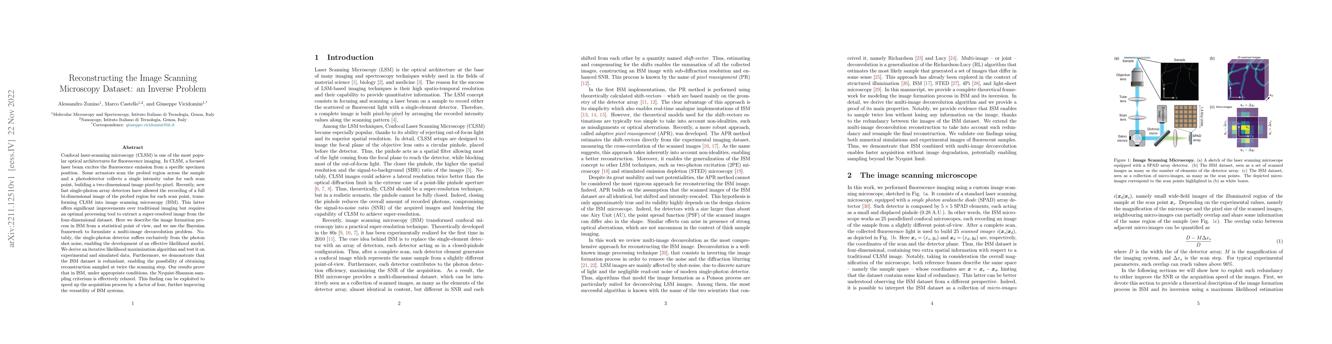
Abstract
Confocal laser-scanning microscopy (CLSM) is one of the most popular optical architectures for fluorescence imaging. In CLSM, a focused laser beam excites the fluorescence emission from a specific specimen position. Some actuators scan the probed region across the sample and a photodetector collects a single intensity value for each scan point, building a two-dimensional image pixel-by-pixel. Recently, new fast single-photon array detectors have allowed the recording of a full bi-dimensional image of the probed region for each scan point, transforming CLSM into image scanning microscopy (ISM). This latter offers significant improvements over traditional imaging but requires an optimal processing tool to extract a super-resolved image from the four-dimensional dataset. Here we describe the image formation process in ISM from a statistical point of view, and we use the Bayesian framework to formulate a multi-image deconvolution problem. Notably, the single-photon detector suffers exclusively from the photon shot noise, enabling the development of an effective likelihood model. We derive an iterative likelihood maximization algorithm and test it on experimental and simulated data. Furthermore, we demonstrate that the ISM dataset is redundant, enabling the possibility of obtaining reconstruction sampled at twice the scanning step. Our results prove that in ISM, under appropriate conditions, the Nyquist-Shannon sampling criterium is effectively relaxed. This finding can be exploited to speed up the acquisition process by a factor of four, further improving the versatility of ISM systems.
AI Key Findings
Get AI-generated insights about this paper's methodology, results, significance, and more — seven facets brought into focus.
Impact
Paper Details
Authors
PDF Preview
Key Terms
Citation Network
Current paper (gray), citations (green), references (blue)
Display is limited for performance on very large graphs.
Discussion 0