Reconstructing the normal and shape at specularities in endoscopy
Publication
Metrics
AI Quick Summary
This paper proposes a novel method to reconstruct tissue normal directions and shape at specularities in endoscopic images, using them as cues for 3D perception rather than treating them as nuisances. Results demonstrate the method's effectiveness on both simulated and real interventional images.
Paper Preview
Abstract
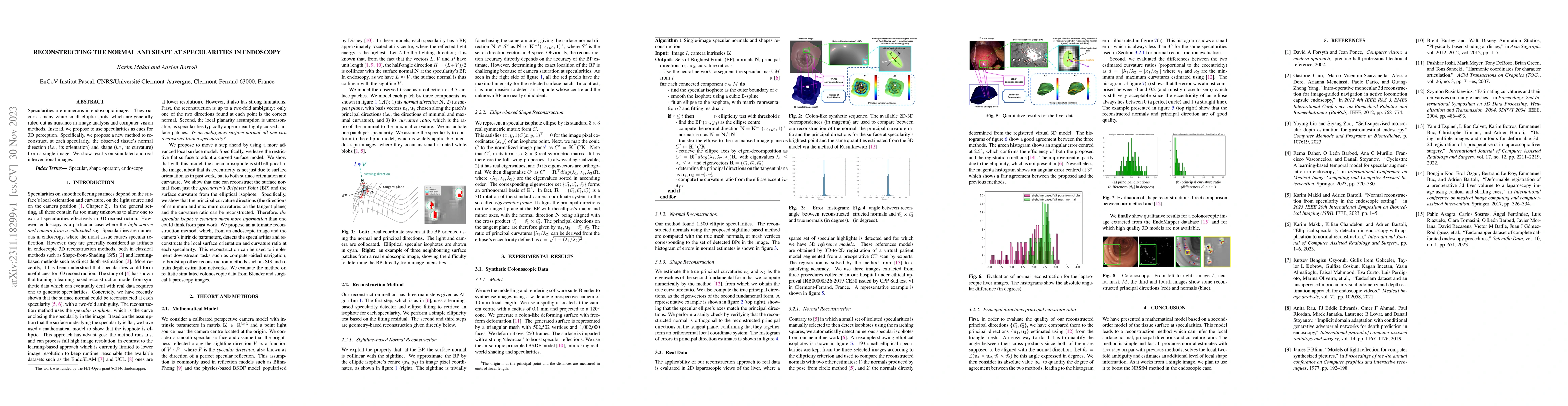
Specularities are numerous in endoscopic images. They occur as many white small elliptic spots, which are generally ruled out as nuisance in image analysis and computer vision methods. Instead, we propose to use specularities as cues for 3D perception. Specifically, we propose a new method to reconstruct, at each specularity, the observed tissue's normal direction (i.e., its orientation) and shape (i.e., its curvature) from a single image. We show results on simulated and real interventional images.
AI Key Findings
Get AI-generated insights about this paper's methodology, results, significance, and more — seven facets brought into focus.
Impact
Paper Details
Authors
PDF Preview
Key Terms
Citation Network
Current paper (gray), citations (green), references (blue)
Display is limited for performance on very large graphs.
Discussion 0