Reconstruction and Quantification of 3D Iris Surface for Angle-Closure Glaucoma Detection in Anterior Segment OCT
Publication
Metrics
AI Quick Summary
This paper proposes a novel framework for reconstructing and quantifying 3D iris surfaces from Anterior Segment OCT (AS-OCT) imagery to detect angle-closure glaucoma, demonstrating that 3D representation provides better performance than 2D methods. The method involves an iris segmentation network with wavelet refinement and guided optimization for 3D reconstruction.
Paper Preview
Abstract
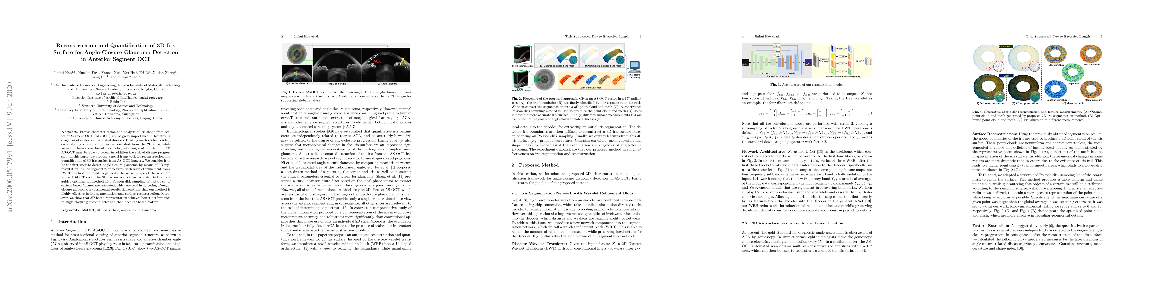
Precise characterization and analysis of iris shape from Anterior Segment OCT (AS-OCT) are of great importance in facilitating diagnosis of angle-closure-related diseases. Existing methods focus solely on analyzing structural properties identified from the 2D slice, while accurate characterization of morphological changes of iris shape in 3D AS-OCT may be able to reveal in addition the risk of disease progression. In this paper, we propose a novel framework for reconstruction and quantification of 3D iris surface from AS-OCT imagery. We consider it to be the first work to detect angle-closure glaucoma by means of 3D representation. An iris segmentation network with wavelet refinement block (WRB) is first proposed to generate the initial shape of the iris from single AS-OCT slice. The 3D iris surface is then reconstructed using a guided optimization method with Poisson-disk sampling. Finally, a set of surface-based features are extracted, which are used in detecting of angle-closure glaucoma. Experimental results demonstrate that our method is highly effective in iris segmentation and surface reconstruction. Moreover, we show that 3D-based representation achieves better performance in angle-closure glaucoma detection than does 2D-based feature.
AI Key Findings
Get AI-generated insights about this paper's methodology, results, significance, and more — seven facets brought into focus.
Impact
Paper Details
Authors
PDF Preview
Key Terms
Citation Network
Current paper (gray), citations (green), references (blue)
Display is limited for performance on very large graphs.
Discussion 0