Recurrent Fully Convolutional Neural Networks for Multi-slice MRI Cardiac Segmentation
Publication
Metrics
AI Quick Summary
This paper proposes a recurrent fully convolutional network (RFCN) for automatic segmentation of the heart in multi-slice MRI, leveraging inter-slice spatial dependencies for improved accuracy. The RFCN architecture combines anatomical detection and segmentation into a single, end-to-end trained model, achieving state-of-the-art results and enhancing contour delineation near the heart's apex.
Paper Preview
Abstract
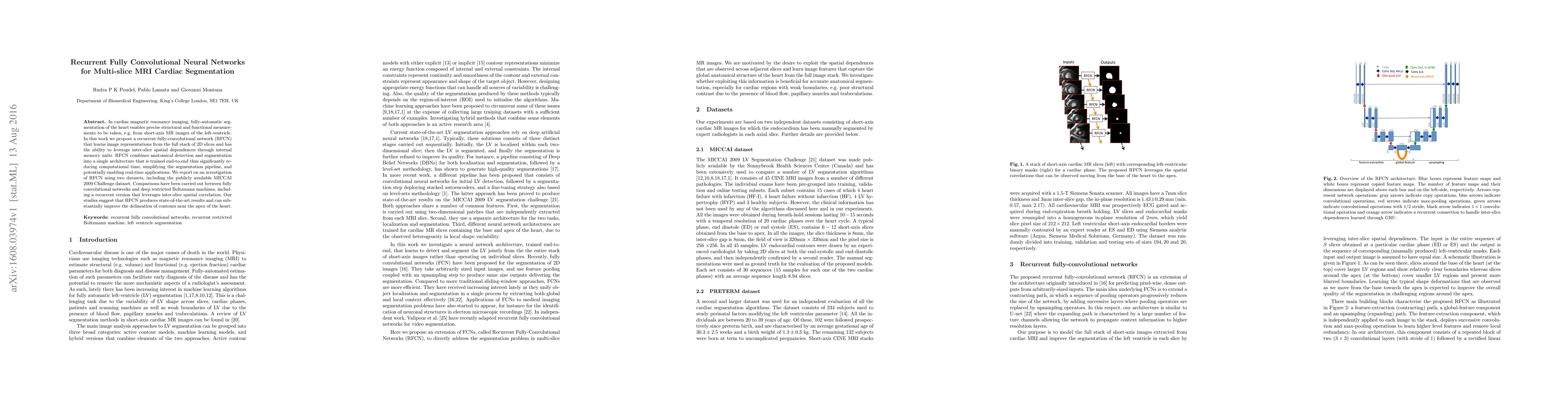
In cardiac magnetic resonance imaging, fully-automatic segmentation of the heart enables precise structural and functional measurements to be taken, e.g. from short-axis MR images of the left-ventricle. In this work we propose a recurrent fully-convolutional network (RFCN) that learns image representations from the full stack of 2D slices and has the ability to leverage inter-slice spatial dependences through internal memory units. RFCN combines anatomical detection and segmentation into a single architecture that is trained end-to-end thus significantly reducing computational time, simplifying the segmentation pipeline, and potentially enabling real-time applications. We report on an investigation of RFCN using two datasets, including the publicly available MICCAI 2009 Challenge dataset. Comparisons have been carried out between fully convolutional networks and deep restricted Boltzmann machines, including a recurrent version that leverages inter-slice spatial correlation. Our studies suggest that RFCN produces state-of-the-art results and can substantially improve the delineation of contours near the apex of the heart.
AI Key Findings
Get AI-generated insights about this paper's methodology, results, significance, and more — seven facets brought into focus.
Impact
Paper Details
PDF Preview
Key Terms
Citation Network
Current paper (gray), citations (green), references (blue)
Display is limited for performance on very large graphs.
Discussion 0