Reducing Shape-Graph Complexity with Application to Classification of Retinal Blood Vessels and Neurons
Publication
Metrics
AI Quick Summary
This paper proposes a method to simplify complex shape graphs through hierarchical clustering, applying it to retinal blood vessel and neuron graphs to retain essential structures. While reduced complexity impairs retinal vessel disease detection, it preserves accuracy for neuron classification.
Paper Preview
Abstract
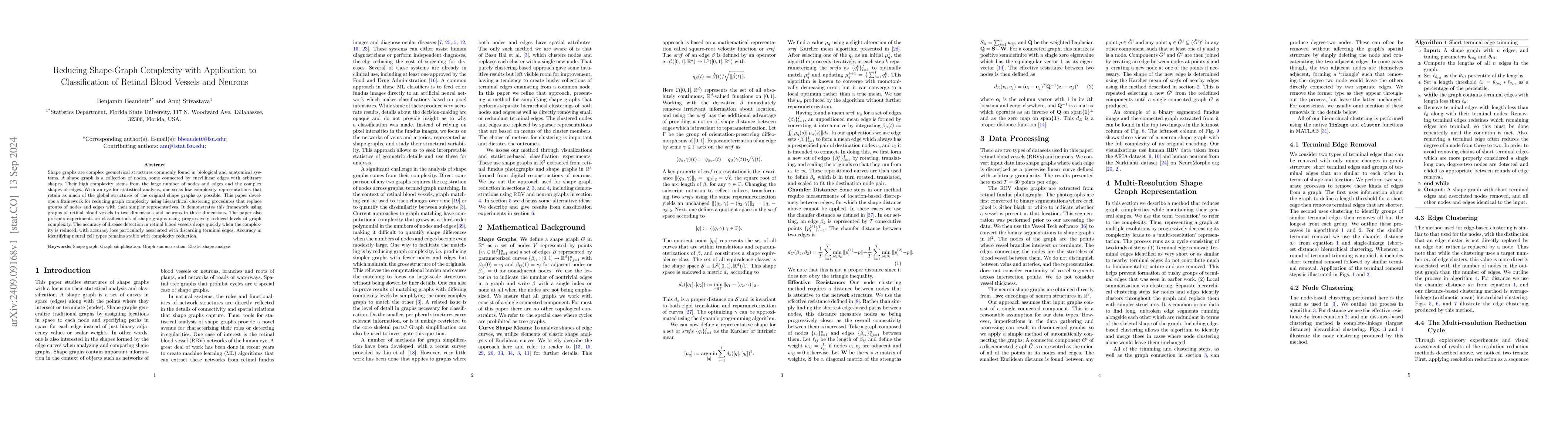
Shape graphs are complex geometrical structures commonly found in biological and anatomical systems. A shape graph is a collection of nodes, some connected by curvilinear edges with arbitrary shapes. Their high complexity stems from the large number of nodes and edges and the complex shapes of edges. With an eye for statistical analysis, one seeks low-complexity representations that retain as much of the global structures of the original shape graphs as possible. This paper develops a framework for reducing graph complexity using hierarchical clustering procedures that replace groups of nodes and edges with their simpler representatives. It demonstrates this framework using graphs of retinal blood vessels in two dimensions and neurons in three dimensions. The paper also presents experiments on classifications of shape graphs using progressively reduced levels of graph complexity. The accuracy of disease detection in retinal blood vessels drops quickly when the complexity is reduced, with accuracy loss particularly associated with discarding terminal edges. Accuracy in identifying neural cell types remains stable with complexity reduction.
AI Key Findings
Get AI-generated insights about this paper's methodology, results, significance, and more — seven facets brought into focus.
Impact
Paper Details
Authors
PDF Preview
Citation Network
Current paper (gray), citations (green), references (blue)
Display is limited for performance on very large graphs.
Discussion 0