Publication
Metrics
AI Quick Summary
This study demonstrates that grating interferometry computed tomography (GI-CT) is more dose-efficient than conventional CT for breast imaging, achieving superior contrast between adipose and glandular tissues. The proposed system, utilizing a 70kVp X-ray tube and commercially available gratings, significantly reduces the required dose while maintaining high image quality, suggesting a promising future for refraction-based imaging in clinical settings.
Paper Preview
Abstract
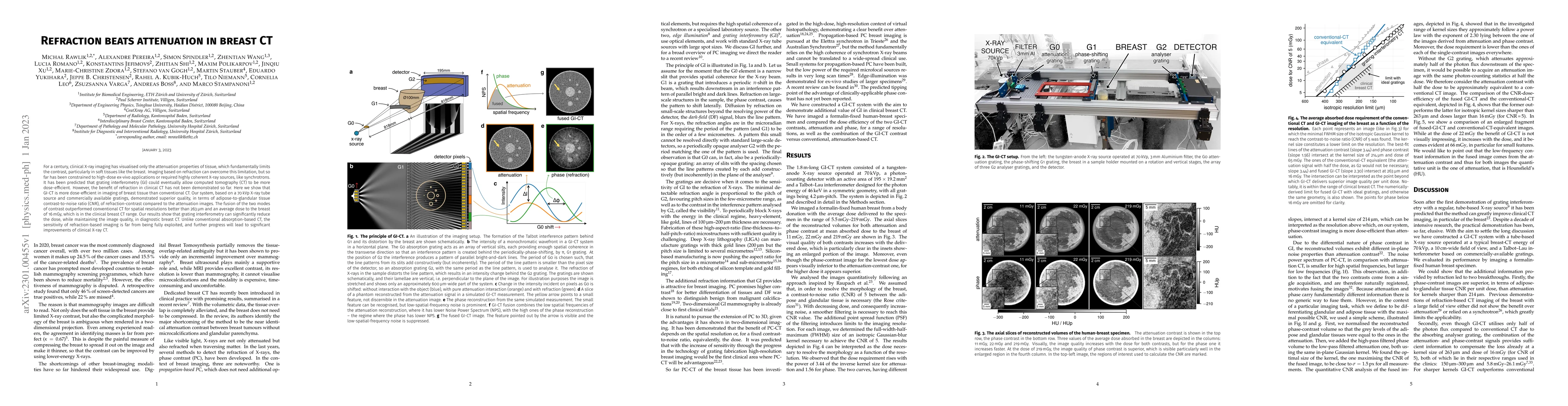
For a century, clinical X-ray imaging has visualised only the attenuation properties of tissue, which fundamentally limits the contrast, particularly in soft tissues like the breast. Imaging based on refraction can overcome this limitation, but so far has been constrained to high-dose ex-vivo applications or required highly coherent X-ray sources, like synchrotrons. It has been predicted that grating interferometry (GI) could eventually allow computed tomography (CT) to be more dose-efficient. However, the benefit of refraction in clinical CT has not been demonstrated so far. Here we show that GI-CT is more dose-efficient in imaging of breast tissue than conventional CT. Our system, based on a 70kVp X-ray tube source and commercially available gratings, demonstrated superior quality, in terms of adipose-to-glandular tissue contrast-to-noise ratio (CNR), of refraction-contrast compared to the attenuation images. The fusion of the two modes of contrast outperformed conventional CT for spatial resolutions better than 263{\mu}m and an average dose to the breast of 16mGy, which is in the clinical breast CT range. Our results show that grating interferometry can significantly reduce the dose, while maintaining the image quality, in diagnostic breast CT. Unlike conventional absorption-based CT, the sensitivity of refraction-based imaging is far from being fully exploited, and further progress will lead to significant improvements of clinical X-ray CT.
AI Key Findings
Get AI-generated insights about this paper's methodology, results, significance, and more — seven facets brought into focus.
Impact
Paper Details
Authors
PDF Preview
Key Terms
Citation Network
Current paper (gray), citations (green), references (blue)
Display is limited for performance on very large graphs.
Discussion 0