01
MethodologyHow they did it
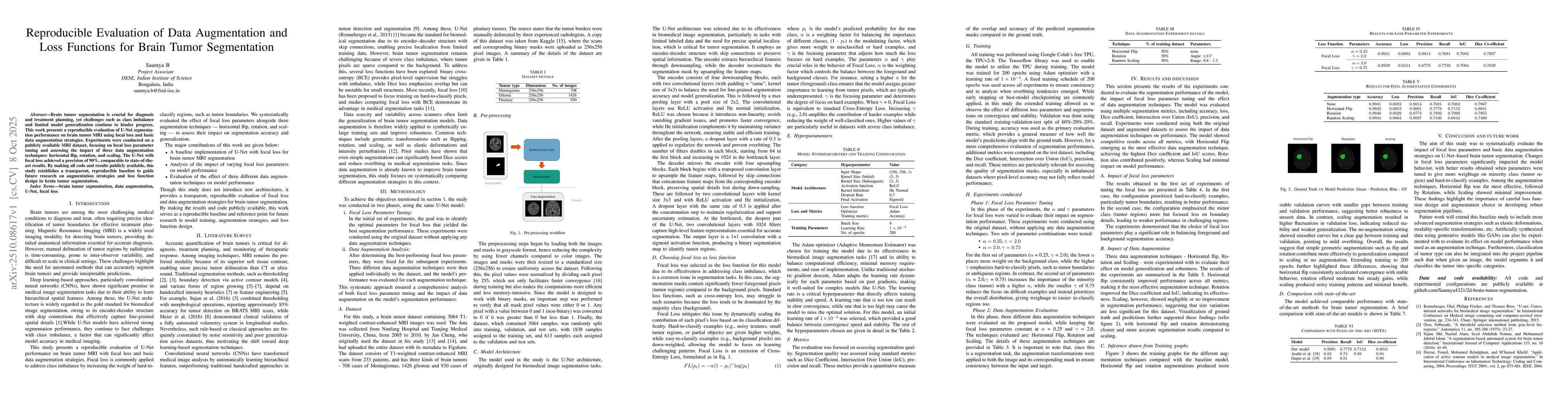
The study employed a two-phase methodology using the U-Net architecture. Phase 1 focused on tuning focal loss parameters (α and γ) on the original dataset without augmentation. Phase 2 evaluated three data augmentation techniques (horizontal flip, rotation, scaling) while keeping focal loss parameters fixed at α=0.25 and γ=2.0.
Discussion 0