Authors
Summary
Chest X-ray (CXR) imaging remains one of the most widely used diagnostic tools for detecting pulmonary diseases such as tuberculosis (TB) and pneumonia. Recent advances in deep learning, particularly Vision Transformers (ViTs), have shown strong potential for automated medical image analysis. However, most ViT architectures are pretrained on natural images and require three-channel inputs, while CXR scans are inherently grayscale. To address this gap, we propose RepViT-CXR, a channel replication strategy that adapts single-channel CXR images into a ViT-compatible format without introducing additional information loss. We evaluate RepViT-CXR on three benchmark datasets. On the TB-CXR dataset,our method achieved an accuracy of 99.9% and an AUC of 99.9%, surpassing prior state-of-the-art methods such as Topo-CXR (99.3% accuracy, 99.8% AUC). For the Pediatric Pneumonia dataset, RepViT-CXR obtained 99.0% accuracy, with 99.2% recall, 99.3% precision, and an AUC of 99.0%, outperforming strong baselines including DCNN and VGG16. On the Shenzhen TB dataset, our approach achieved 91.1% accuracy and an AUC of 91.2%, marking a performance improvement over previously reported CNN-based methods. These results demonstrate that a simple yet effective channel replication strategy allows ViTs to fully leverage their representational power on grayscale medical imaging tasks. RepViT-CXR establishes a new state of the art for TB and pneumonia detection from chest X-rays, showing strong potential for deployment in real-world clinical screening systems.
AI Key Findings
Generated Nov 02, 2025
Methodology
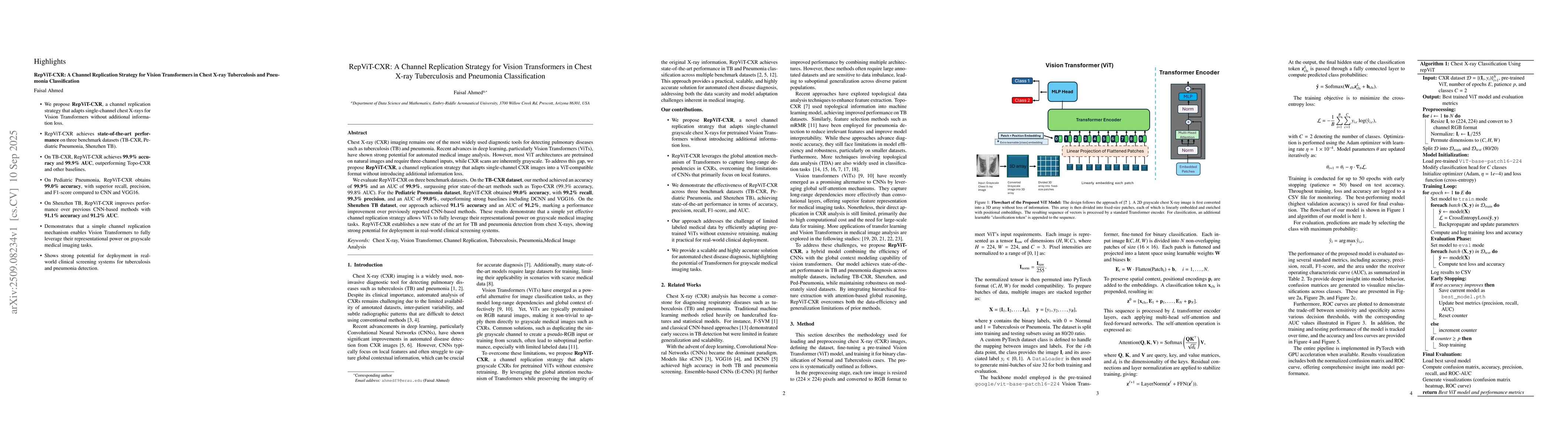
The research proposes RepViT-CXR, a channel replication strategy that adapts single-channel CXR images into a ViT-compatible format by converting grayscale images into 3-channel RGB format without information loss. The model uses a pre-trained ViT-base-patch16-224 architecture, fine-tuned for binary classification, with data split into 80/20 training/test sets.
Key Results
- RepViT-CXR achieved 99.9% accuracy and 99.9% AUC on TB-CXR dataset, surpassing prior state-of-the-art methods like Topo-CXR (99.3% accuracy, 99.8% AUC)
- On Pediatric Pneumonia dataset, RepViT-CXR obtained 99.0% accuracy with 99.2% recall, 99.3% precision, and 99.0% AUC, outperforming DCNN and VGG16
- On Shenzhen TB dataset, RepViT-CXR achieved 91.1% accuracy and 91.2% AUC, improving upon CNN-based methods
Significance
This research establishes a new state-of-the-art for TB and pneumonia detection from chest X-rays, demonstrating ViTs' potential in grayscale medical imaging tasks and enabling reliable automated diagnosis for clinical screening systems.
Technical Contribution
Developed a channel replication strategy to adapt grayscale CXR images for ViT architectures without information loss, enabling effective use of pre-trained ViT weights for medical image analysis.
Novelty
Introduces a simple yet effective channel replication strategy that allows ViTs to leverage their representational power on grayscale medical imaging tasks, bridging the gap between natural image-based ViTs and medical imaging applications.
Limitations
- Channel replication does not add new information, it only ensures compatibility with pre-trained ViTs
- Performance on Shenzhen TB dataset showed lower AUC (91.2) compared to ResNet-BS (95.4)
Future Work
- Explore modality-aware embeddings or self-supervised pretraining strategies tailored to medical images
- Investigate domain adaptation techniques to handle variations due to demographic, scanner, or clinical setting differences
Paper Details
PDF Preview
Similar Papers
Found 4 papersDINO-CXR: A self supervised method based on vision transformer for chest X-ray classification
Mahdi Eftekhari, Mohammadreza Shakouri, Fatemeh Iranmanesh
Deep transfer learning for detecting Covid-19, Pneumonia and Tuberculosis using CXR images -- A Review
Kinyua Gikunda, Irad Mwendo, Anthony Maina
Comments (0)