Resolution enhancement of placenta histological images using deep learning
Publication
Metrics
AI Quick Summary
This study developed a deep learning method to enhance the resolution of histological placenta images using a modified U-net model trained on paired high- and low-resolution images. The method achieved a relative mean squared error of 0.003, improving image contrast and adding critical details.
Paper Preview
Abstract
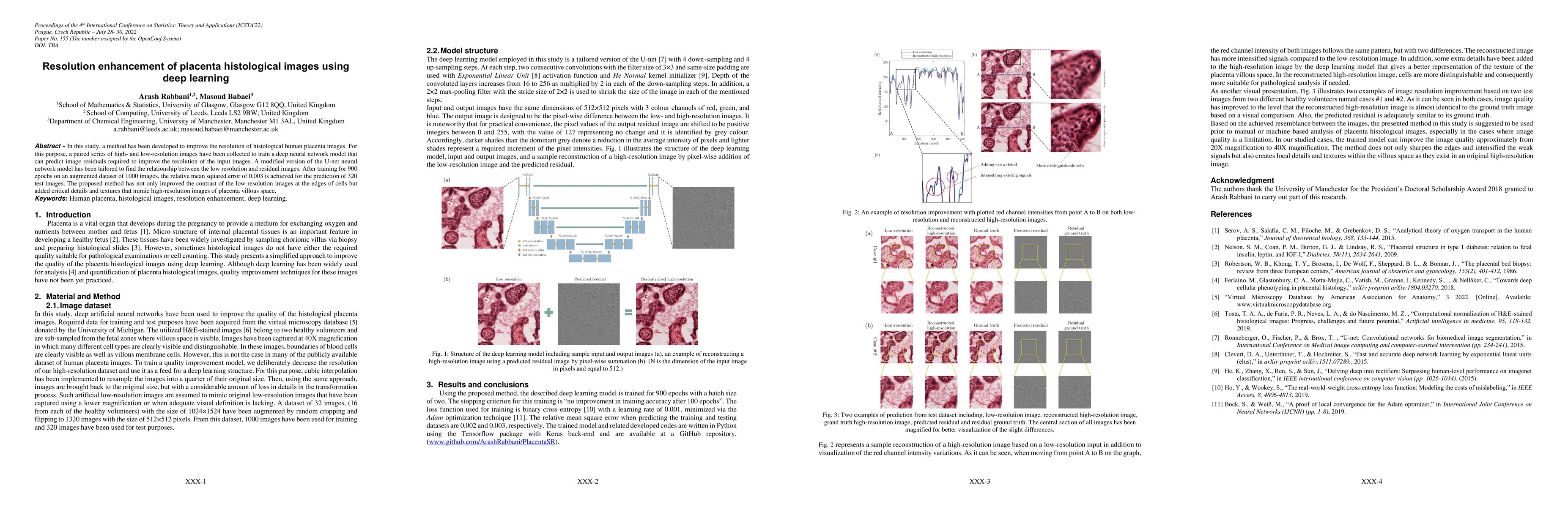
In this study, a method has been developed to improve the resolution of histological human placenta images. For this purpose, a paired series of high- and low-resolution images have been collected to train a deep neural network model that can predict image residuals required to improve the resolution of the input images. A modified version of the U-net neural network model has been tailored to find the relationship between the low resolution and residual images. After training for 900 epochs on an augmented dataset of 1000 images, the relative mean squared error of 0.003 is achieved for the prediction of 320 test images. The proposed method has not only improved the contrast of the low-resolution images at the edges of cells but added critical details and textures that mimic high-resolution images of placenta villous space.
AI Key Findings
Get AI-generated insights about this paper's methodology, results, significance, and more — seven facets brought into focus.
Impact
Paper Details
Authors
PDF Preview
Key Terms
Citation Network
Current paper (gray), citations (green), references (blue)
Display is limited for performance on very large graphs.
Discussion 0