Resolution Enhancement of Under-sampled Photoacoustic Microscopy Images using Implicit Neural Representations
Publication
Metrics
AI Quick Summary
The paper proposes a method based on Implicit Neural Representations to enhance the resolution of under-sampled Photoacoustic Microscopy images, addressing limitations of traditional interpolation and deconvolution techniques. This approach improves resolution and reduces scanning times, demonstrated through significant PSNR and SSIM gains on simulated and real vascular data.
Paper Preview
Abstract
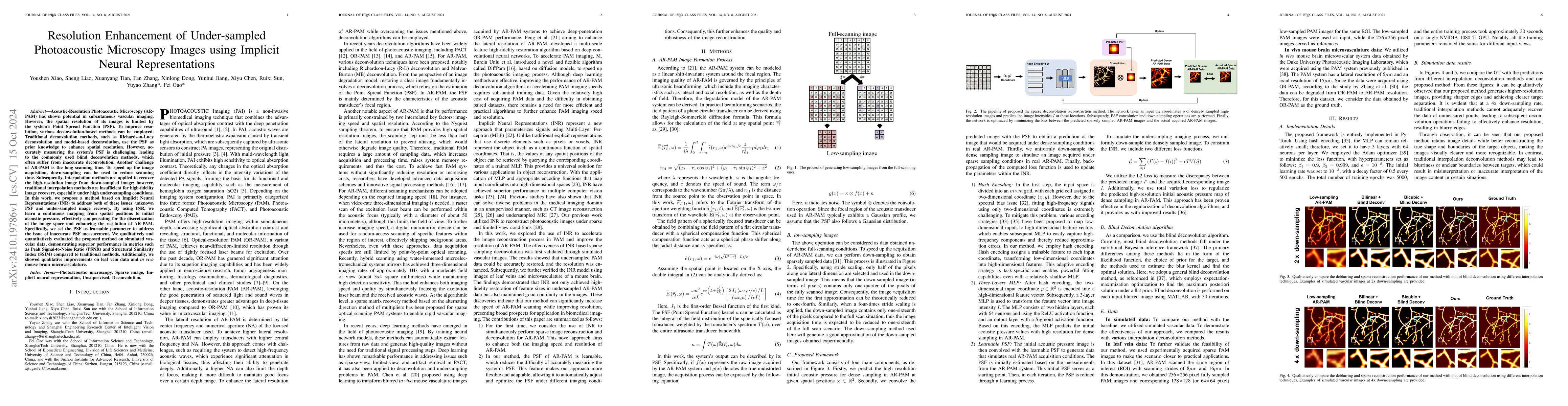
Acoustic-Resolution Photoacoustic Microscopy (AR-PAM) is promising for subcutaneous vascular imaging, but its spatial resolution is constrained by the Point Spread Function (PSF). Traditional deconvolution methods like Richardson-Lucy and model-based deconvolution use the PSF to improve resolution. However, accurately measuring the PSF is difficult, leading to reliance on less accurate blind deconvolution techniques. Additionally, AR-PAM suffers from long scanning times, which can be reduced via down-sampling, but this necessitates effective image recovery from under-sampled data, a task where traditional interpolation methods fall short, particularly at high under-sampling rates. To address these challenges, we propose an approach based on Implicit Neural Representations (INR). This method learns a continuous mapping from spatial coordinates to initial acoustic pressure, overcoming the limitations of discrete imaging and enhancing AR-PAM's resolution. By treating the PSF as a learnable parameter within the INR framework, our technique mitigates inaccuracies associated with PSF estimation. We evaluated our method on simulated vascular data, showing significant improvements in Peak Signal-to-Noise Ratio (PSNR) and Structural Similarity Index (SSIM) over conventional methods. Qualitative enhancements were also observed in leaf vein and in vivo mouse brain microvasculature images. When applied to a custom AR-PAM system, experiments with pencil lead demonstrated that our method delivers sharper, higher-resolution results, indicating its potential to advance photoacoustic microscopy.
AI Key Findings
Get AI-generated insights about this paper's methodology, results, significance, and more — seven facets brought into focus.
Impact
Authors
PDF Preview
Citation Network
Current paper (gray), citations (green), references (blue)
Display is limited for performance on very large graphs.
Discussion 0