Summary

Self-supervised learning (SSL) has advanced visual representation learning, but its value in chest radiography, a high-volume imaging modality with fine-grained findings, remains unclear. Meta's DINOv3 extends earlier SSL models through Gram-anchored self-distillation. Whether these design choices improve transfer learning for chest radiography has not been systematically 
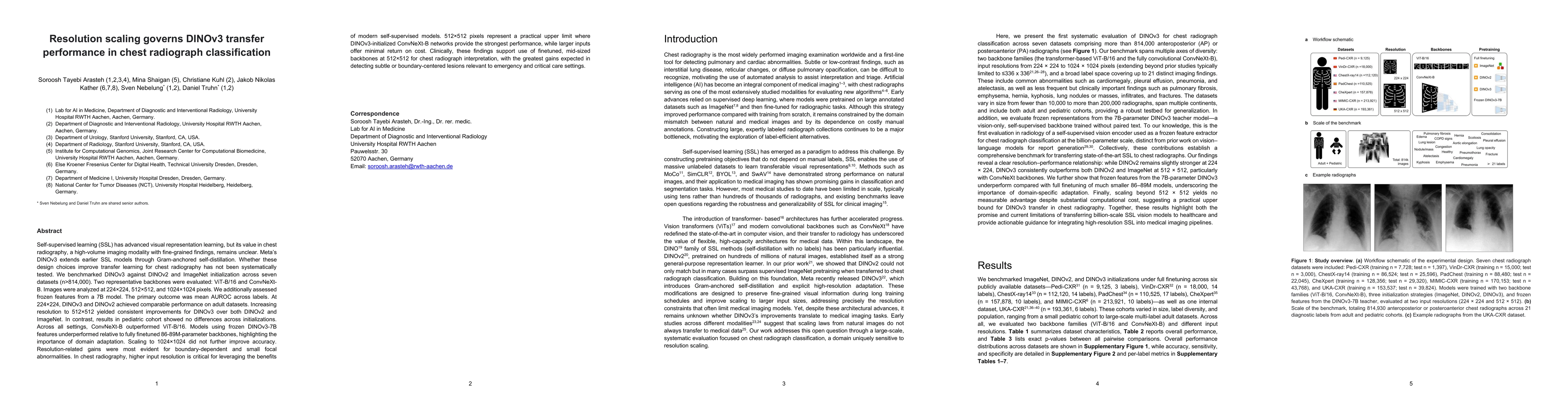
tested. We benchmarked DINOv3 against DINOv2 and ImageNet initialization across seven datasets (n>814,000). Two representative backbones were evaluated: ViT-B/16 and ConvNeXt-B. Images were analyzed at 224x224, 512x512, and 1024x1024 pixels. We additionally assessed frozen features from a 7B model. The primary outcome was mean AUROC across labels. At 224x224, DINOv3 and DINOv2 achieved comparable performance on adult datasets. Increasing resolution to 512x512 yielded consistent improvements for DINOv3 over both DINOv2 and ImageNet. In contrast, results in pediatric cohort showed no differences across initializations. Across all settings, ConvNeXt-B outperformed ViT-B/16. Models using frozen DINOv3-7B features underperformed relative to fully finetuned 86-89M-parameter backbones, highlighting the importance of domain adaptation. Scaling to 1024x1024 did not further improve accuracy. Resolution-related gains were most evident for boundary-dependent and small focal abnormalities. In chest radiography, higher input resolution is critical for leveraging the benefits of modern self-supervised models. 512x512 pixels represent a practical upper limit where DINOv3-initialized ConvNeXt-B networks provide the strongest performance, while larger inputs offer minimal return on cost. Clinically, these findings support use of finetuned, mid-sized backbones at 512x512 for chest radiograph interpretation, with the greatest gains expected in detecting subtle or boundary-centered lesions relevant to emergency and critical care settings.

AI Key Findings

Generated Oct 10, 2025

Methodology

The study compared different initialization strategies (ImageNet, DINOv2, DINOv3) for pre-trained models (ViT-B/16 and ConvNeXt-B) across multiple chest X-ray datasets. Performance was evaluated using AUROC, accuracy, sensitivity, and specificity metrics through full fine-tuning and feature extraction approaches.

Key Results

  • DINOv3 outperformed ImageNet and DINOv2 across most datasets and resolutions
  • Full fine-tuning of smaller models consistently outperformed frozen DINOv3-7B features
  • Performance improvements from DINOv3 increased with higher resolution in multi-label datasets

Significance

This research advances medical image analysis by demonstrating the effectiveness of self-supervised pre-training for chest X-ray diagnosis. The findings could improve diagnostic accuracy and reduce reliance on labeled data in clinical settings.

Technical Contribution

The study provides comprehensive benchmarking of different pre-training methods for chest X-ray analysis, offering insights into optimal initialization strategies and model architectures for medical imaging tasks.

Novelty

This work is novel in systematically comparing DINOv2 and DINOv3 pre-training methods for chest X-ray analysis, and in demonstrating the benefits of high-resolution feature extraction for multi-label medical image classification.

Limitations

  • Results are primarily based on adult datasets with limited pediatric data
  • Performance metrics may vary with different clinical populations and imaging protocols

Future Work

  • Investigate DINOv3's effectiveness in other medical imaging modalities
  • Explore hybrid training approaches combining self-supervised and supervised learning
  • Validate results in real-world clinical environments

Paper Details

PDF Preview

Download PDF

Comments (0)

This website uses cookies to ensure you get the best experience. Learn more