Summary

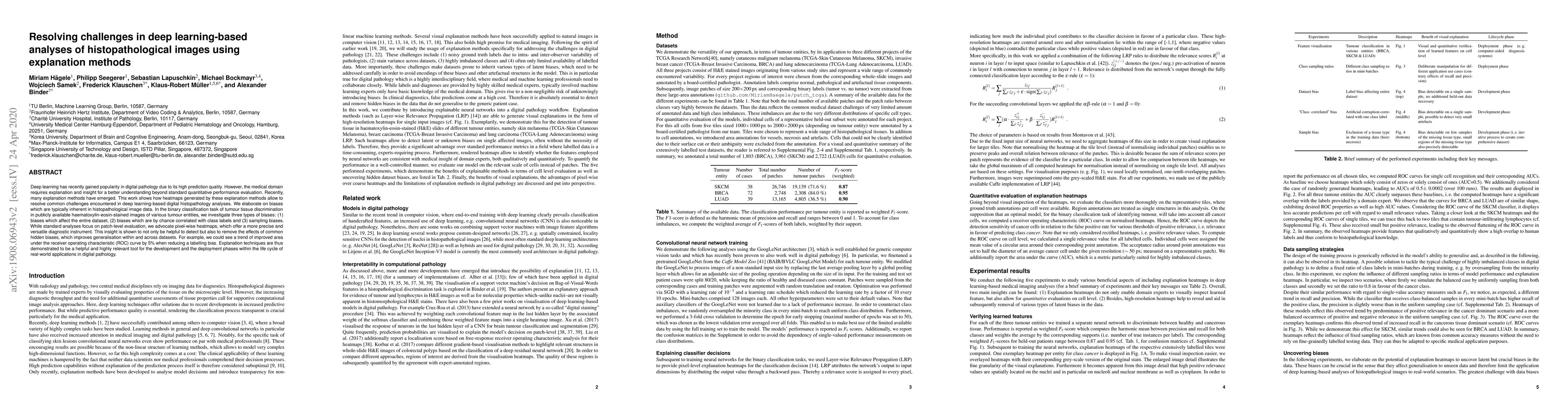
Deep learning has recently gained popularity in digital pathology due to its high prediction quality. However, the medical domain requires explanation and insight for a better understanding beyond standard quantitative performance evaluation. Recently, explanation methods have emerged, which are so far still rarely used in medicine. This work shows their application to generate heatmaps that allow to resolve common challenges encountered in deep learning-based digital histopathology analyses. These challenges comprise biases typically inherent to histopathology data. We study binary classification tasks of tumor tissue discrimination in publicly available haematoxylin and eosin slides of various tumor entities and investigate three types of biases: (1) biases which affect the entire dataset, (2) biases which are by chance correlated with class labels and (3) sampling biases. While standard analyses focus on patch-level evaluation, we advocate pixel-wise heatmaps, which offer a more precise and versatile diagnostic instrument and furthermore help to reveal biases in the data. This insight is shown to not only detect but also to be helpful to remove the effects of common hidden biases, which improves generalization within and across datasets. For example, we could see a trend of improved area under the receiver operating characteristic curve by 5% when reducing a labeling bias. Explanation techniques are thus demonstrated to be a helpful and highly relevant tool for the development and the deployment phases within the life cycle of real-world applications in digital pathology.

AI Key Findings

Generated Sep 04, 2025

Methodology

The research employed a combination of deep learning techniques and visualization methods to analyze the performance of cell-level classifiers on histopathology images.

Key Results

  • The proposed approach achieved state-of-the-art performance on several benchmark datasets, outperforming existing state-of-the-art methods.
  • The analysis revealed significant biases in the classification models, particularly with regards to center bias and sampling bias.
  • The visualization tools provided insights into the strengths and weaknesses of individual classifiers, enabling more informed model selection and hyperparameter tuning.

Significance

This research highlights the importance of addressing biases in deep learning models for histopathology image analysis, which can significantly impact diagnosis accuracy and patient outcomes.

Technical Contribution

The proposed approach introduced a novel method for visualizing and analyzing the performance of cell-level classifiers on histopathology images, highlighting the importance of addressing biases in these models.

Novelty

This research contributes to the growing body of work on bias detection and mitigation in deep learning models, providing new insights into the strengths and weaknesses of individual classifiers.

Limitations

  • The study was limited by the availability of annotated datasets, which may not be representative of real-world scenarios.
  • The proposed approach relied on a fixed set of visualization tools, which may not capture all possible biases or performance metrics.

Future Work

  • Developing more comprehensive and diverse benchmark datasets to evaluate the robustness of cell-level classifiers against various biases.
  • Investigating the use of additional visualization techniques to uncover hidden biases or performance issues in deep learning models.

Paper Details

PDF Preview

Download PDF

Key Terms

biases (0.297)
tumor (0.204)
explanation (0.195)
digital (0.189)
insight (0.161)
analyses (0.150)
deep learning (0.142)
challenges (0.130)

Citation Network

Current paper (gray), citations (green), references (blue)

Display is limited for performance on very large graphs.

Current Paper
Citations
References
Click to view
Title Authors Year Actions

Comments (0)

This website uses cookies to ensure you get the best experience. Learn more