Summary

In this study, we curated a densely annotated in-house dataset comprising 490 CTPA scans. Using this dataset, we systematically evaluated nine widely used segmentation architectures from both the CNN and Vision Transformer (ViT) families, initialized with either pretrained or random weights, under a unified testing framework as a performance audit. Our study leads to several important observations: (1) 3D U-Net with a ResNet encoder remains a highly effective architecture for PE segmentation; (2) 3D models are particularly well-suited to this task given the morphological characteristics of emboli; (3) CNN-based models generally yield superior performance compared to their ViT-based counterparts in PE segmentation; (4) classification-based pretraining, even on large PE datasets, can adversely impact segmentation performance compared to training from scratch, suggesting that PE classification and segmentation may rely on different sets of discriminative features; (5) different model architectures show a highly consistent pattern of segmentation performance when trained on the same data; and (6) while central and large emboli can be segmented with satisfactory accuracy, distal emboli remain challenging due to both task complexity and the scarcity of high-quality datasets. Besides these 
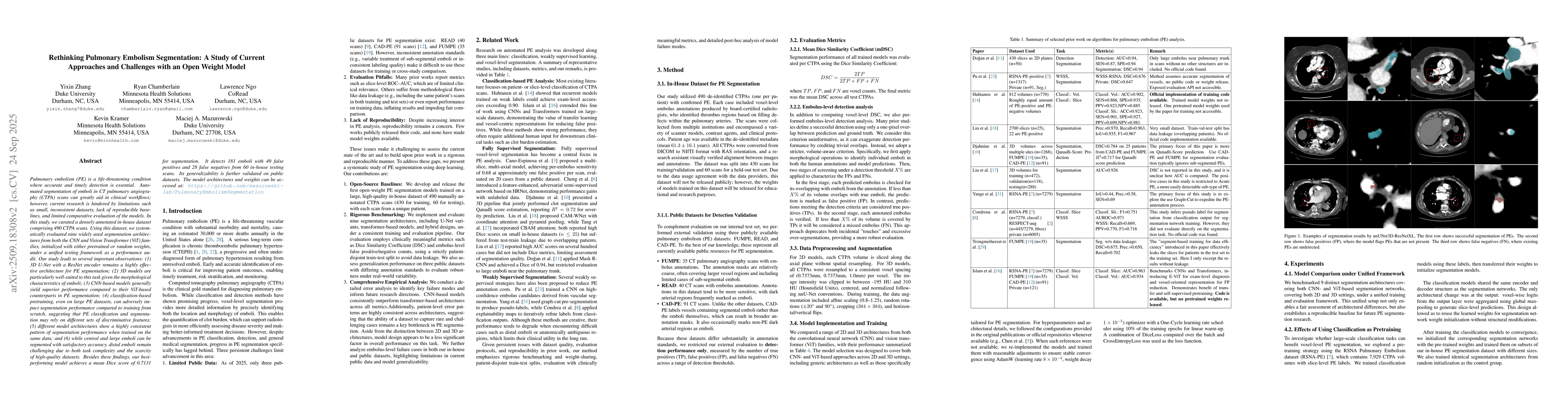
findings, our best-performing model achieves a mean Dice score of 0.7131 for segmentation. It detects 181 emboli with 49 false positives and 28 false negatives from 60 in-house testing scans. Its generalizability is further validated on public datasets.

AI Key Findings

Generated Sep 29, 2025

Methodology

The study curates a dataset of 490 CTPA scans and evaluates nine segmentation architectures (CNN and ViT-based) using a unified testing framework. Models are initialized with pretrained or random weights, and performance is assessed across multiple metrics including Dice score, sensitivity, and specificity.

Key Results

  • 3D U-Net with ResNet encoder remains highly effective for PE segmentation
  • CNN-based models outperform ViT-based models in PE segmentation
  • Distal emboli are challenging due to task complexity and limited high-quality data

Significance

This research provides a comprehensive evaluation of PE segmentation methods, identifies effective architectures, and highlights critical challenges in distal embolus detection, offering guidance for future model development and dataset creation.

Technical Contribution

Introduces the first set of open-weight baselines for PE segmentation and establishes a reproducible benchmarking framework for future research.

Novelty

Combines systematic evaluation of CNN and ViT architectures with analysis of pretraining impacts, while providing open weights for direct use in medical imaging tasks.

Limitations

  • Annotations in public datasets may have inconsistencies due to varying annotation protocols
  • Pretraining on classification tasks did not improve segmentation performance compared to random initialization

Future Work

  • Develop larger and clinically validated datasets for small/distal PE
  • Create data-leakage-free evaluation protocols reflecting clinical usability
  • Prioritize reproducibility through code and pretrained weight releases

Paper Details

PDF Preview

Download PDF

Comments (0)

This website uses cookies to ensure you get the best experience. Learn more