Retinal blood flow speed quantification at the capillary level using temporal autocorrelation fitting OCTA
Publication
Metrics
AI Quick Summary
This paper introduces a second-generation OCTA technique, VISTA, to quantify blood flow speed at the capillary level using temporal autocorrelation fitting. The method employs a 600 kHz A-scan rate for high-resolution retinal imaging and demonstrates the cardiac pulsatility and repeatability of the flow speed marker, showing different flow speeds in various retinal capillary plexuses.
Paper Preview
Abstract
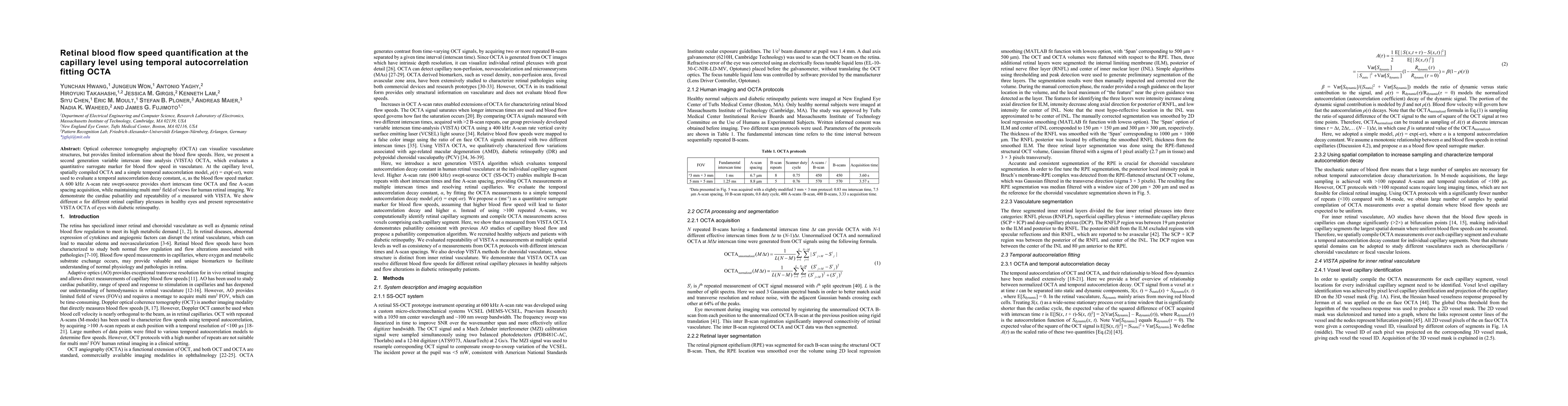
Optical coherence tomography angiography (OCTA) can visualize vasculature structures, but provides limited information about the blood flow speeds. Here, we present a second generation variable interscan time analysis (VISTA) OCTA, which evaluates a quantitative surrogate marker for blood flow speed in vasculature. At the capillary level, spatially compiled OCTA and a simple temporal autocorrelation model, {\rho}({\tau}) = exp(-{\alpha}{\tau}), were used to evaluate a temporal autocorrelation decay constant, {\alpha}, as the blood flow speed marker. A 600 kHz A-scan rate swept-source provides short interscan time OCTA and fine A-scan spacing acquisition, while maintaining multi mm2 field of views for human retinal imaging. We demonstrate the cardiac pulsatility and repeatability of {\alpha} measured with VISTA. We show different {\alpha} for different retinal capillary plexuses in healthy eyes and present representative VISTA OCTA of eyes with diabetic retinopathy.
AI Key Findings
Get AI-generated insights about this paper's methodology, results, significance, and more — seven facets brought into focus.
Impact
Paper Details
Authors
PDF Preview
Key Terms
Citation Network
Current paper (gray), citations (green), references (blue)
Display is limited for performance on very large graphs.
Discussion 0