Retrieval-augmented Few-shot Medical Image Segmentation with Foundation Models
Publication
Metrics
AI Quick Summary
This paper introduces a retrieval-augmented few-shot medical image segmentation method leveraging DINOv2 and Segment Anything Model 2 (SAM 2), which uses memory attention to generate accurate segmentations without requiring retraining or finetuning, demonstrating superior performance across multiple modalities.
Paper Preview
Abstract
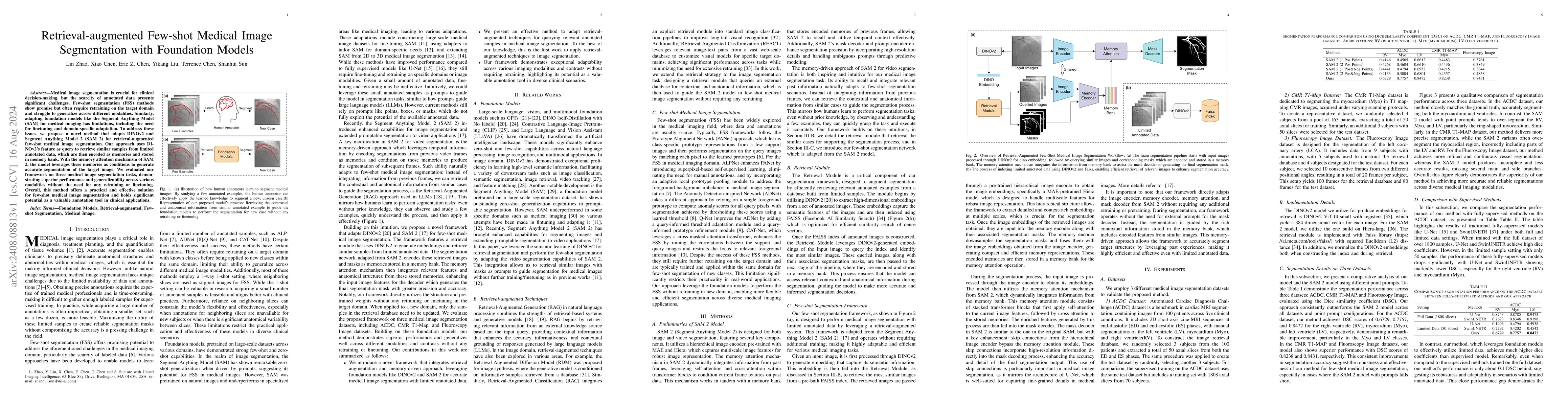
Medical image segmentation is crucial for clinical decision-making, but the scarcity of annotated data presents significant challenges. Few-shot segmentation (FSS) methods show promise but often require retraining on the target domain and struggle to generalize across different modalities. Similarly, adapting foundation models like the Segment Anything Model (SAM) for medical imaging has limitations, including the need for finetuning and domain-specific adaptation. To address these issues, we propose a novel method that adapts DINOv2 and Segment Anything Model 2 (SAM 2) for retrieval-augmented few-shot medical image segmentation. Our approach uses DINOv2's feature as query to retrieve similar samples from limited annotated data, which are then encoded as memories and stored in memory bank. With the memory attention mechanism of SAM 2, the model leverages these memories as conditions to generate accurate segmentation of the target image. We evaluated our framework on three medical image segmentation tasks, demonstrating superior performance and generalizability across various modalities without the need for any retraining or finetuning. Overall, this method offers a practical and effective solution for few-shot medical image segmentation and holds significant potential as a valuable annotation tool in clinical applications.
AI Key Findings
Get AI-generated insights about this paper's methodology, results, significance, and more — seven facets brought into focus.
Impact
Authors
PDF Preview
Key Terms
Citation Network
Current paper (gray), citations (green), references (blue)
Display is limited for performance on very large graphs.
Discussion 0