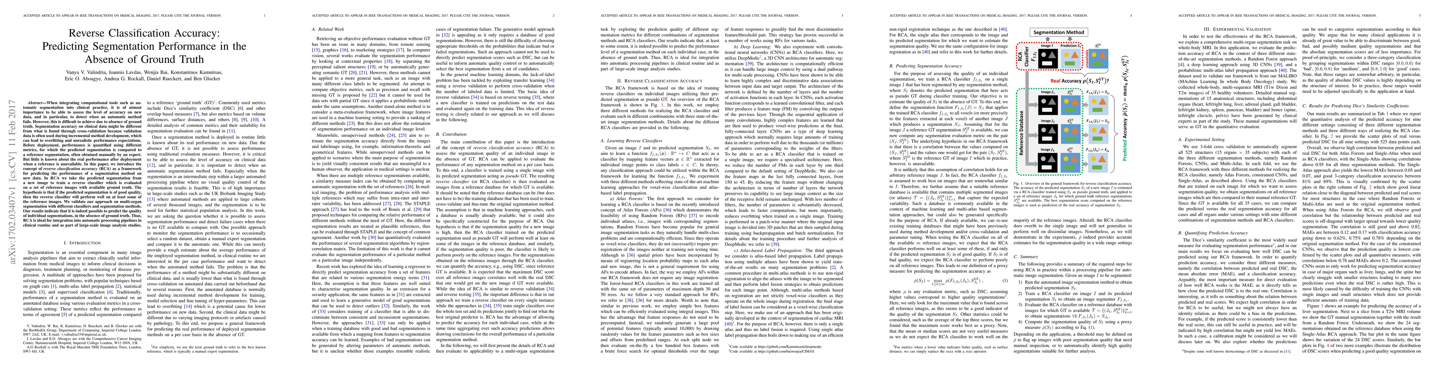
When integrating computational tools such as automatic segmentation into
clinical practice, it is of utmost importance to be able to assess the level of
accuracy on new data, and in particular, to detect when an automatic method
fails. However, this is difficult to achieve due to absence of ground truth.
Segmentation accuracy on clinical data might be different from what is found
through cross-validation because validation data is often used during
incremental method development, which can lead to overfitting and unrealistic
performance expectations. Before deployment, performance is quantified using
different metrics, for which the predicted segmentation is compared to a
reference segmentation, often obtained manually by an expert. But little is
known about the real performance after deployment when a reference is
unavailable. In this paper, we introduce the concept of reverse classification
accuracy (RCA) as a framework for predicting the performance of a segmentation
method on new data. In RCA we take the predicted segmentation from a new image
to train a reverse classifier which is evaluated on a set of reference images
with available ground truth. The hypothesis is that if the predicted
segmentation is of good quality, then the reverse classifier will perform well
on at least some of the reference images. We validate our approach on
multi-organ segmentation with different classifiers and segmentation methods.
Our results indicate that it is indeed possible to predict the quality of
individual segmentations, in the absence of ground truth. Thus, RCA is ideal
for integration into automatic processing pipelines in clinical routine and as
part of large-scale image analysis studies.
Discussion 0