Publication
Metrics
AI Quick Summary
This paper presents a robust diffusion imaging framework designed to handle artefacts in clinical studies, employing an improved least trimmed squares diffusion tensor estimation algorithm to restore corrupted datasets. The framework's efficiency and accuracy are demonstrated through simulations and in vivo experiments, showing its potential for broader use in MR studies requiring artefact suppression.
Paper Preview
Abstract
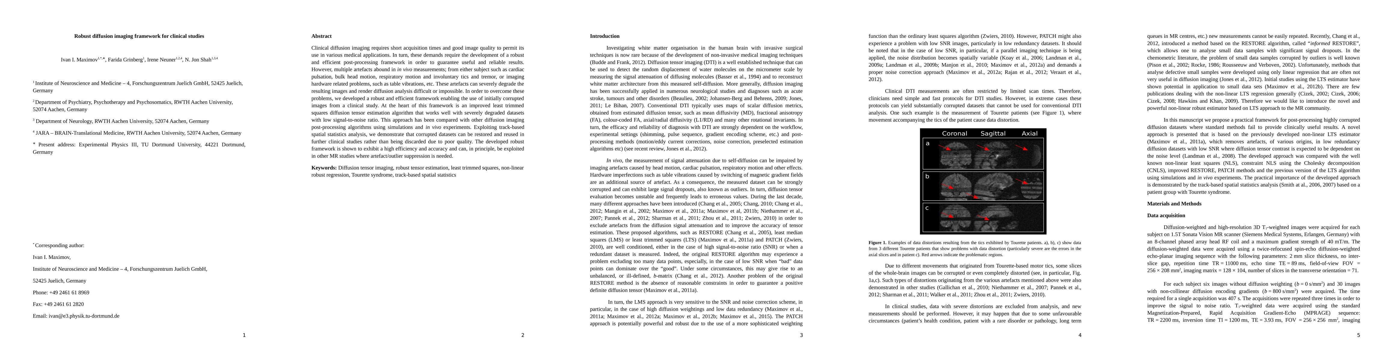
Clinical diffusion imaging requires short acquisition times and good image quality to permit its use in various medical applications. In turn, these demands require the development of a robust and efficient post-processing framework in order to guarantee useful and reliable results. However, multiple artefacts abound in in vivo measurements; from either subject such as cardiac pulsation, bulk head motion, respiratory motion and involuntary tics and tremor, or imaging hardware related problems, such as table vibrations, etc. These artefacts can severely degrade the resulting images and render diffusion analysis difficult or impossible. In order to overcome these problems, we developed a robust and efficient framework enabling the use of initially corrupted images from a clinical study. At the heart of this framework is an improved least trimmed squares diffusion tensor estimation algorithm that works well with severely degraded datasets with low signal-to-noise ratio. This approach has been compared with other diffusion imaging post-processing algorithms using simulations and in vivo experiments. Exploiting track-based spatial statistics analysis, we demonstrate that corrupted datasets can be restored and reused in further clinical studies rather than being discarded due to poor quality. The developed robust framework is shown to exhibit a high efficiency and accuracy and can, in principle, be exploited in other MR studies where artefact/outlier suppression is needed.
AI Key Findings
Get AI-generated insights about this paper's methodology, results, significance, and more — seven facets brought into focus.
Impact
Paper Details
PDF Preview
Key Terms
Citation Network
Current paper (gray), citations (green), references (blue)
Display is limited for performance on very large graphs.
Discussion 0