ArxivLens

Quick Summary:

This paper proposes a self-learning framework for robust pancreatic ductal adenocarcinoma (PDAC) segmentation using multi-institutional, multi-phase CT scans with limited annotations. The method generates pseudo annotations from two teacher models and refines them with a teaching assistant, achieving a 6.3% Dice score improvement over the baseline.

arXiv Id: 2008.10652
Comments: 10 pages, 2 figures; MICCAI 2020
Date Published: 2020-08-26
Date Updated: 2020-08-26

Categories:

Key Terms (TF-IDF):

annotated (0.294)
annotated (0.276)
segmentation (0.271)
tumor (0.269)
segmentation (0.254)
images (0.196)
ct (0.191)
images (0.188)
annotations (0.180)
annotations (0.177)
patient (0.174)
patient (0.173)
clinical (0.162)
imaging (0.162)
clinical (0.162)
imaging (0.160)
pseudo (0.151)
pseudo (0.150)
trained (0.125)
trained (0.120)
License: http://arxiv.org/licenses/nonexclusive-distrib/1.0/

Summary:

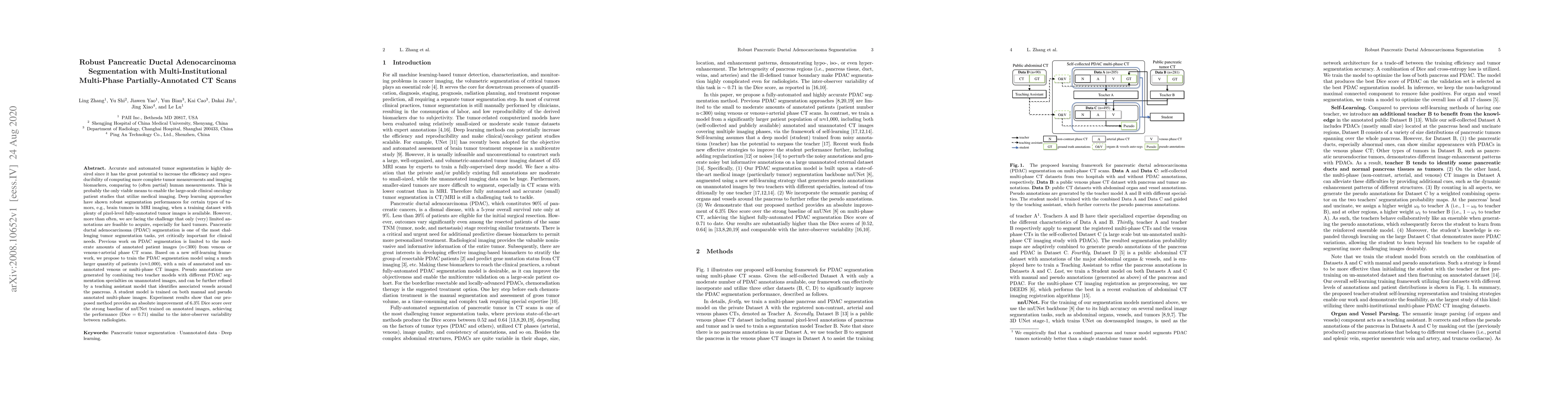
Accurate and automated tumor segmentation is highly desired since it has the great potential to increase the efficiency and reproducibility of computing more complete tumor measurements and imaging biomarkers, comparing to (often partial) human measurements. This is probably the only viable means to enable the large-scale clinical oncology patient studies that utilize medical imaging. Deep learning approaches have shown robust segmentation performances for certain types of tumors, e.g., brain tumors in MRI imaging, when a training dataset with plenty of pixel-level fully-annotated tumor images is available. However, more than often, we are facing the challenge that only (very) limited annotations are feasible to acquire, especially for hard tumors. Pancreatic ductal adenocarcinoma (PDAC) segmentation is one of the most challenging tumor segmentation tasks, yet critically important for clinical needs. Previous work on PDAC segmentation is limited to the moderate amounts of annotated patient images (n<300) from venous or venous+arterial phase CT scans. Based on a new self-learning framework, we propose to train the PDAC segmentation model using a much larger quantity of patients (n~=1,000), with a mix of annotated and un-annotated venous or multi-phase CT images. Pseudo annotations are generated by combining two teacher models with different PDAC segmentation specialties on unannotated images, and can be further refined by a teaching assistant model that identifies associated vessels around the pancreas. A student model is trained on both manual and pseudo annotated multi-phase images. Experiment results show that our proposed method provides an absolute improvement of 6.3% Dice score over the strong baseline of nnUNet trained on annotated images, achieving the performance (Dice = 0.71) similar to the inter-observer variability between radiologists.

Citation & Reference Network Visualization

This graph shows how this paper is connected to other research. The current paper is shown in dark gray, papers that cite it are in green, and papers it references are in blue. You can:

Current Paper
Papers citing this work (Citations)
Papers cited by this work (References)

• Hover over any node to see the paper title

• Click a node to view that paper's details

• Drag nodes to rearrange the graph

• Use the zoom controls (bottom right) to zoom in/out

Citations and References

Title Authors Year Actions
Evidence modeling for reliability learning and interpretable decision-making under multi-modality medical image segmentation
Jianfeng Zhao, Shuo Li
2024
View Paper
Optimizing Universal Lesion Segmentation: State Space Model-Guided Hierarchical Networks with Feature Importance Adjustment
Kazi Shahriar Sanjid, Md. Tanzim Hossain, Md. Shakib Shahariar Junayed, M. M. Uddin
2024
View Paper
Artificial intelligence for assessment of vascular involvement and tumor resectability on CT in patients with pancreatic cancer
Jacqueline I Bereska, Boris V. Janssen, C. Y. Nio, Marnix P. M. Kop, G. Kazemier, O. Busch, F. Struik, Henk A. Marquering, Jaap Stoker, M. Besselink, I. Verpalen
2024
View Paper
Acquiring Weak Annotations for Tumor Localization in Temporal and Volumetric Data
Yu-Cheng Chou, Bowen Li, Deng-Ping Fan, Alan L. Yuille, Zongwei Zhou
2023
View Paper
Improved Prognostic Prediction of Pancreatic Cancer Using Multi-Phase CT by Integrating Neural Distance and Texture-Aware Transformer
Hexin Dong, Jiawen Yao, Yuxing Tang, Ming Yuan, Yingda Xia, Jian Zhou, Hong Lu, Jing Zhou, Bin Dong, Le Lu, Li Zhang, Zai-De Liu, Yu Shi, Ling Zhang
2023
View Paper
Meta-information-aware Dual-path Transformer for Differential Diagnosis of Multi-type Pancreatic Lesions in Multi-phase CT
Bo Zhou, Yingda Xia, Jiawen Yao, Le Lu, Jingren Zhou, Chi Liu, J. Duncan, Ling Zhang
2023
View Paper
The FELIX Project: Deep Networks To Detect Pancreatic Neoplasms
Y. Xia, Q. Yu, L. Chu, S. Kawamoto, S. Park, F. Liu, J. Chen, Z. Zhu, B. Li, Z. Zhou, Y. Lu, Y. Wang, W. Shen, L. Xie, Y. Zhou, elliot k fishman, A. Javed, D. Fouladi, S. Shayesteh, J. Graves, A. Blanco, E. Zinreich, B. Kinny-Koster, K. Kinzler, R. Hruban, B. Vogelstein, A. Yuille, E. Fishman
2022
View Paper
External Attention Assisted Multi-Phase Splenic Vascular Injury Segmentation With Limited Data
Yuyin Zhou, D. Dreizin, Yan Wang, Fengze Liu, Wei-lei Shen, A. Yuille
2021
View Paper
Detect-and-Segment: a Deep Learning Approach to Automate Wound Image Segmentation
G. Scebba, Jia Zhang, Sabrina Catanzaro, C. Mihai, O. Distler, M. Berli, W. Karlen
2021
View Paper
Clinical Artificial Intelligence Applications in Radiology: Chest and Abdomen.
Sungwon Lee, Ronald M. Summers
2021
View Paper
DeepStationing: Thoracic Lymph Node Station Parsing in CT Scans using Anatomical Context Encoding and Key Organ Auto-Search
Dazhou Guo, X. Ye, J. Ge, Xing Di, Le Lu, Lingyun Huang, G. Xie, Jing Xiao, Zhongjie Liu, Ling Peng, Senxiang Yan, D. Jin
2021
View Paper
DeepPrognosis: Preoperative prediction of pancreatic cancer survival and surgical margin via comprehensive understanding of dynamic contrast-enhanced CT imaging and tumor-vascular contact parsing
Jiawen Yao, Yu-long Shi, K. Cao, Le Lu, Jianping Lu, Qike Song, G. Jin, Jing Xiao, Yang Hou, Ling Zhang
2021
View Paper
Segmentation of pancreatic ductal adenocarcinoma (PDAC) and surrounding vessels in CT images using deep convolutional neural networks and texture descriptors
Tahereh Mahmoudi, Zahra Mousavi Kouzahkanan, A. Radmard, R. Kafieh, Aneseh Salehnia, A. Davarpanah, H. Arabalibeik, A. Ahmadian
2021
View Paper
Weakly-Supervised Universal Lesion Segmentation with Regional Level Set Loss
Youbao Tang, Jinzheng Cai, K. Yan, Lingyun Huang, G. Xie, Jing Xiao, Jingjing Lu, Gigin Lin, Le Lu
2021
View Paper
Deep Lesion Tracker: Monitoring Lesions in 4D Longitudinal Imaging Studies
Jinzheng Cai, Youbao Tang, K. Yan, Adam P. Harrison, Jing Xiao, Gigin Lin, Le Lu
2020
View Paper
3D Graph Anatomy Geometry-Integrated Network for Pancreatic Mass Segmentation, Diagnosis, and Quantitative Patient Management
Tianyi Zhao, Kai Cao, Jiawen Yao, Isabella Nogues, Le Lu, Lingyun Huang, Jing Xiao, Zhaozheng Yin, Ling Zhang
2020
View Paper
U-Net-based Pancreas Tumor Segmentation from Abdominal CT Images
H. S. Saraswathi, M. Rafi
2023
View Paper
A Teacher-Student Framework with Fourier Augmentation for COVID-19 Infection Segmentation in CT Images
Han Chen, Yifan Jiang, Hanseok Ko, Murray H. Loew
2021
View Paper
Effective Pancreatic Cancer Screening on Non-contrast CT Scans via Anatomy-Aware Transformers
Yingda Xia, Jiawen Yao, Le Lu, Lingyun Huang, G. Xie, Jing Xiao, A. Yuille, K. Cao, Ling Zhang
2021
View Paper

Comments (0)

This website uses cookies to ensure you get the best experience. Learn more