ArxivLens

Quick Summary:

This research introduces Re-scan Second Harmonic Generation Microscopy (rSHG) for label-free 3D visualization, achieving resolutions surpassing the diffraction limit by ~1.4x. The study also demonstrates super-resolved Re-scan Two-Photon Excited Fluorescence Microscopy (rTPEF), implemented via a modified Re-scan Confocal Microscope, thus creating a versatile multimodal imaging system.

DOI: 10.1073/pnas.2214662119
arXiv Id: 2207.02002
Date Published: 2022-11-16
Date Updated: 2022-11-16

Key Terms (TF-IDF):

scan (0.364)
scan (0.357)
microscopy (0.330)
microscopy (0.315)
imaging (0.239)
resolved (0.237)
imaging (0.237)
fluorescence (0.236)
super (0.236)
resolved (0.231)
super (0.229)
harmonic (0.189)
harmonic (0.189)
reported (0.174)
reported (0.169)
generation (0.155)
generation (0.150)
optical (0.143)
optical (0.140)
multimodal (0.136)
License: http://creativecommons.org/licenses/by/4.0/

Summary:

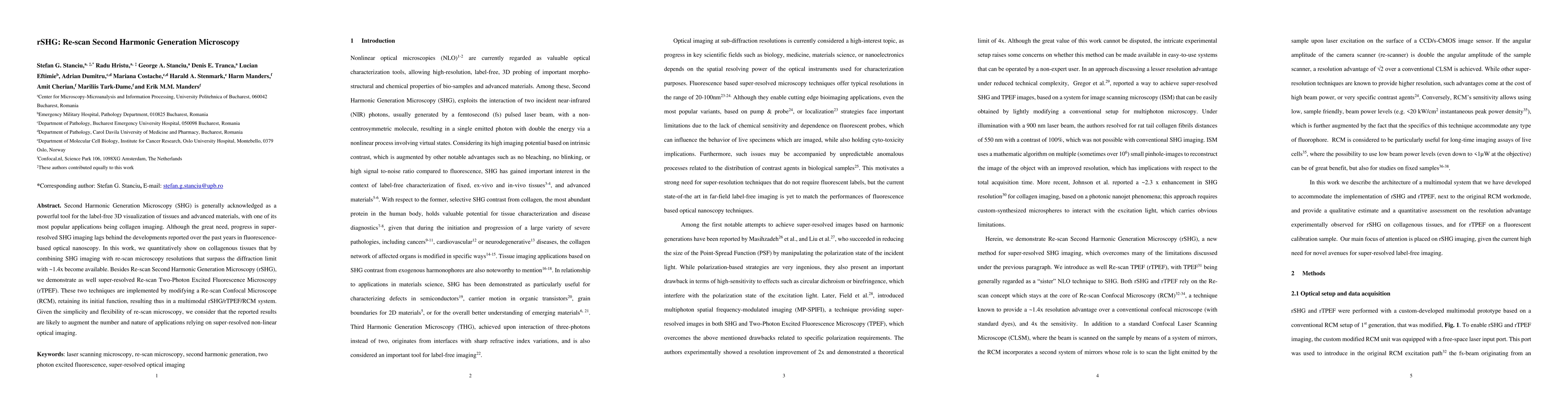
Second Harmonic Generation Microscopy (SHG) is generally acknowledged as a powerful tool for the label-free 3D visualization of tissues and advanced materials, with one of its most popular applications being collagen imaging. Although the great need, progress in super-resolved SHG imaging lags behind the developments reported over the past years in fluorescence-based optical nanoscopy. In this work, we quantitatively show on collagenous tissues that by combining SHG imaging with re-scan microscopy resolutions that surpass the diffraction limit with ~1.4x become available. Besides Re-scan Second Harmonic Generation Microscopy (rSHG), we demonstrate as well super-resolved Re-scan Two-Photon Excited Fluorescence Microscopy (rTPEF). These two techniques are implemented by modifying a Re-scan Confocal Microscope (RCM), retaining its initial function, resulting thus in a multimodal rSHG/rTPEF/RCM system. Given the simplicity and flexibility of re-scan microscopy, we consider that the reported results are likely to augment the number and nature of applications relying on super-resolved non-linear optical imaging.

Citation & Reference Network Visualization

This graph shows how this paper is connected to other research. The current paper is shown in dark gray, papers that cite it are in green, and papers it references are in blue. You can:

Current Paper
Papers citing this work (Citations)
Papers cited by this work (References)

• Hover over any node to see the paper title

• Click a node to view that paper's details

• Drag nodes to rearrange the graph

• Use the zoom controls (bottom right) to zoom in/out

Citations and References

Title Authors Year Actions
Quantitative and comparative assessment of dyes and protocols for rapid ex vivo microscopy of fresh tissues
Ivan Bozic, M. Behr, J. Q. Brown
2024
View Paper
Two-Photon Absorption Aggregation-Induced Emission Luminogen/Paclitaxel Nanoparticles for Cancer Theranostics.
Liwei Zhu, Yiming Wang, Jiayi Song, Zonghai Sheng, Ji Qi, Ying Li, Guoxin Li, B. Tang
2024
View Paper
Widefield polarization-resolved second harmonic generation imaging of entire thyroid nodule sections for the detection of capsular invasion
L. Eftimie, Y. Padrez, L. Golubewa, D. Rutkauskas, R. Hristu
2024
View Paper
Optical Phenomena in Molecule-Based Magnetic Materials
Jakub J. Zakrzewski, Michał Liberka, Junhao Wang, S. Chorazy, S. Ohkoshi
2024
View Paper
Super-resolved coherent anti-Stokes Raman scattering microscopy by coherent image scanning
Anna Zhitnitsky, Elad Benjamin, O. Bitton, D. Oron
2024
View Paper
Modeling and predicting second harmonic generation from protein molecular structure
Bahar Asadipour, Emmanuel Beaurepaire, Xingjian Zhang, A. Chessel, Pierre Mahou, W. Supatto, M. Schanne-Klein, C. Stringari
2024
View Paper
Generic arrays of surface-positioned and shallow-buried gold multi-shapes as reference samples to benchmark near-field microscopes. Part 1: Applications in s-SNOM depth imaging
Binyamin Kusnetz, J. Belhassen, D. Tranca, S. Stanciu, Stefan R. Anton, Z. Zalevsky, G. Stanciu, A. Karsenty
2024
View Paper
Roadmap on Label‐Free Super‐Resolution Imaging
V. Astratov, Yair Ben Sahel, Y. Eldar, Luzhe Huang, Aydogan Ozcan, Nikolay Zheludev, Junxiang Zhao, Zachary Burns, Zhaowei Liu, E. Narimanov, Neha Goswami, Gabriel Popescu, Emanuel Pfitzner, Philipp Kukura, Yi-Teng Hsiao, Chia‐Lung Hsieh, Brian Abbey, A. Diaspro, Aymeric LeGratiet, P. Bianchini, N. Shaked, B. Simon, N. Verrier, M. Debailleul, O. Haeberlé, Sheng Wang, Mengkun Liu, Yeran Bai, Ji‐Xin Cheng, B. Kariman, Katsumasa Fujita, M. Sinvani, Z. Zalevsky, Xiang Li, Guan-Jie Huang, Shi-Wei Chu, Omer Tzang, D. Hershkovitz, O. Cheshnovsky, M. Huttunen, S. Stanciu, V. Smolyaninova, I. Smolyaninov, Ulf Leonhardt, Sahar Sahebdivan, Zengbo Wang, B. Luk’yanchuk, Limin Wu, A. V. Maslov, B. Jin, C. Simovski, S. Perrin, Paul Montgomery, S. Lecler
2023
View Paper
Super-Resolution Second-Harmonic Generation Imaging with Multifocal Structured Illumination Microscopy.
Chenshuang Zhang, Fangrui Lin, Yong Zhang, Haozhi Yang, Danying Lin, Jun He, C. Liao, Xiaoyu Weng, Liwei Liu, Yiping Wang, Bin Yu, Junle Qu
2023
View Paper
AutoFibroNet: A deep learning and multi‐photon microscopy‐derived automated network for liver fibrosis quantification in MAFLD
Huiling Zhan, Siyu Chen, F. Gao, Guangxing Wang, Sui-Dan Chen, G. Xi, Hai-Yang Yuan, Xiaolu Li, Wen-Yue Liu, C. Byrne, G. Targher, Miao-Yang Chen, Yong‐Feng Yang, Jun Chen, Zhiwen Fan, Xitai Sun, Guorong Cai, M. Zheng, S. Zhuo
2023
View Paper
Super-Resolved Non-linear Optical Microscopy: Architectures, Advantages and Perspectives
S. Stanciu, R. Hristu, G. Stanciu, D. Tranca, L. Eftimie, A. Dumitru, M. Costache, H. Stenmark, J. Bueno, P. Bianchini, E. Manders
2023
View Paper
Revealing the Structural Organization of Gamma-irradiated Starch Granules Using Polarization-resolved Second Harmonic Generation Microscopy.
Ming-Chi Chen, Indira Govindaraju, Wei-Hsun Wang, Wei‐Liang Chen, K. D. Mumbrekar, S. Mal, B. Sarmah, V. J. Baruah, P. Srisungsitthisunti, Naregundi Karunakara, N. Mazumder, Guan-Yu Zhuo
2023
View Paper
Toward next-generation endoscopes integrating biomimetic video systems, nonlinear optical microscopy, and deep learning.
S. Stanciu, Karsten König, Young Min Song, L. Wolf, C. Charitidis, P. Bianchini, M. Goetz
2023
View Paper
Viewing life without labels under optical microscopes
Biswajoy Ghosh, Krishna Agarwal
2023
View Paper

Comments (0)

This website uses cookies to ensure you get the best experience. Learn more