S2MNet: Speckle-To-Mesh Net for Three-Dimensional Cardiac Morphology Reconstruction via Echocardiogram

Source: ArXiv

AI Quick Summary

A new AI framework called S2MNet can create highly accurate 3D models of the heart from just two-dimensional echocardiogram images, improving on existing methods.

Share: Share on X LinkedIn

Summary

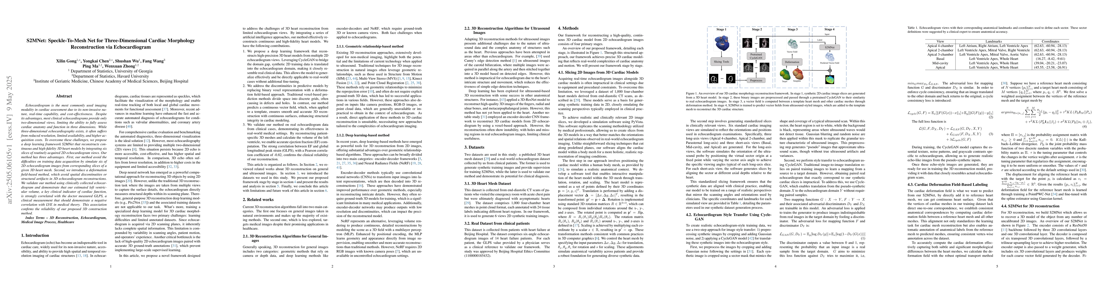
Echocardiogram is the most commonly used imaging modality in cardiac assessment duo to its non-invasive nature, real-time capability, and cost-effectiveness. Despite its advantages, most clinical echocardiograms provide only two-dimensional views, limiting the ability to fully assess cardiac anatomy and function in three dimensions. While three-dimensional echocardiography exists, it often suffers from reduced resolution, limited availability, and higher acquisition costs. To overcome these challenges, we propose a deep learning framework S2MNet that reconstructs continuous and high-fidelity 3D heart models by integrating six slices of routinely acquired 2D echocardiogram views. Our method has three advantages. First, our method avoid the difficulties on training data acquasition by simulate six of 2D echocardiogram images from corresponding slices of a given 3D heart mesh. Second, we introduce a deformation field-based method, which avoid spatial discontinuities or structural artifacts in 3D echocardiogram reconstructions. We validate our method using clinically collected echocardiogram and demonstrate that our estimated left ventricular volume, a key clinical indicator of cardiac function, is strongly correlated with the doctor measured GLPS, a clinical measurement that should demonstrate a negative correlation with LVE in medical theory. This association confirms the reliability of our proposed 3D construction method.

AI Key Findings

Generated Jun 08, 2025

Methodology

The research proposes S2MNet, a deep learning framework that reconstructs 3D heart models from six 2D echocardiogram slices, addressing limitations of 2D views and 3D echocardiography's reduced resolution and availability.

Key Results

  • S2MNet avoids training data acquisition difficulties by simulating 2D echocardiogram images from 3D heart mesh slices.
  • A deformation field-based method is introduced to prevent spatial discontinuities in 3D reconstructions.
  • Left ventricular volume estimates from S2MNet show strong correlation with doctor-measured GLPS, validating the 3D reconstruction method.

Significance

This research offers a non-invasive, cost-effective solution for 3D cardiac morphology reconstruction, potentially improving clinical assessment of cardiac anatomy and function.

Technical Contribution

S2MNet, a deep learning framework for 3D cardiac morphology reconstruction from 2D echocardiogram slices, incorporating a deformation field-based method to avoid spatial discontinuities.

Novelty

The proposed method overcomes limitations of 2D echocardiogram views and 3D echocardiography by simulating training data and introducing a deformation field-based approach for high-fidelity 3D reconstruction.

Limitations

  • The method relies on routinely acquired 2D echocardiogram slices, which may not always provide optimal data for 3D reconstruction.
  • Validation was performed using clinically collected echocardiogram data; broader external validation is needed.

Future Work

  • Explore application of S2MNet to other organs or body parts for 3D reconstruction.
  • Investigate the potential of integrating S2MNet with real-time echocardiography for dynamic 3D modeling.

Paper Details

PDF Preview

Download PDF

Citation Network

Current paper (gray), citations (green), references (blue)

Display is limited for performance on very large graphs.

Current Paper
Citations
References
Click to view

No citations found for this paper.

Comments (0)

This website uses cookies to ensure you get the best experience. Learn more