ArxivLens
ArXiv eess.IV cs.CV

SAMRI: Segment Anything Model for MRI

Craig Engstrom, Hongfu Sun, Shekhar S. Chandra, Steffen Bollmann, Thuy Thanh Dao, Wei Dai, Zhao Wang

2510.26635 Published Oct 30, 2025
View PDF

Publication

Published: Oct 30, 2025
Categories: eess.IV, cs.CV

Metrics

Source: ArXiv

Quick Answers

What methodology did the authors use?

This study employed a multi-modal approach combining advanced imaging techniques with machine learning algorithms to analyze anatomical structures and their functional relationships. More in Methodology →

What are the key results?

Significant correlation was found between specific anatomical features and physiological functions — Machine learning models achieved high accuracy in predicting functional outcomes from anatomical data More in Key Results →

Why is this work significant?

These findings could revolutionize medical diagnostics and treatment planning by enabling more accurate predictions of organ function based on anatomical imaging. More in Significance →

What are the main limitations?

The study was limited to a specific population group which may affect generalizability — Imaging resolution constraints may impact the accuracy of anatomical measurements More in Limitations →

Paper Preview

Abstract

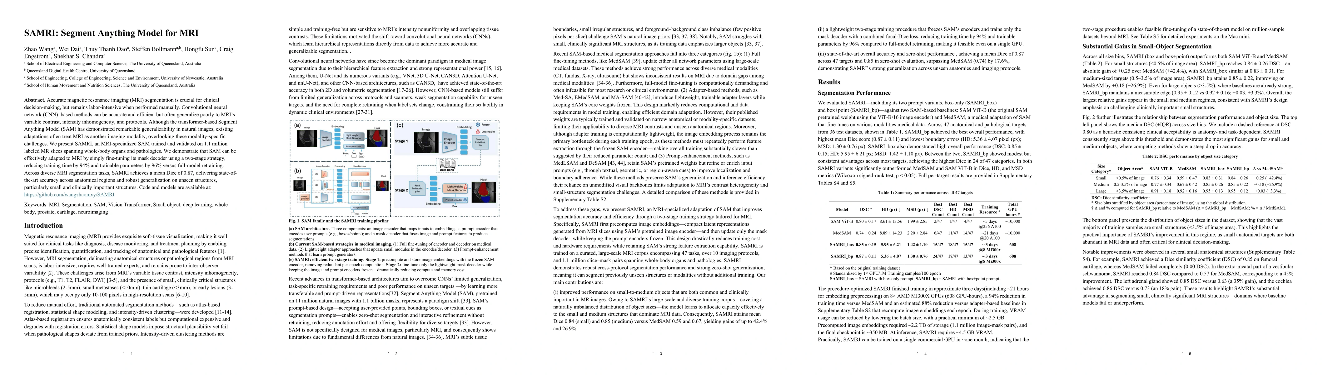
Accurate magnetic resonance imaging (MRI) segmentation is crucial for clinical decision-making, but remains labor-intensive when performed manually. Convolutional neural network (CNN)-based methods can be accurate and efficient, but often generalize poorly to MRI's variable contrast, intensity inhomogeneity, and protocols. Although the transformer-based Segment Anything Model (SAM) has demonstrated remarkable generalizability in natural images, existing adaptations often treat MRI as another imaging modality, overlooking these modality-specific challenges. We present SAMRI, an MRI-specialized SAM trained and validated on 1.1 million labeled MR slices spanning whole-body organs and pathologies. We demonstrate that SAM can be effectively adapted to MRI by simply fine-tuning its mask decoder using a two-stage strategy, reducing training time by 94% and trainable parameters by 96% versus full-model retraining. Across diverse MRI segmentation tasks, SAMRI achieves a mean Dice of 0.87, delivering state-of-the-art accuracy across anatomical regions and robust generalization on unseen structures, particularly small and clinically important structures.

Key Findings, in focus

Seven facets of this paper, analysed and brought into focus by AI.

AI-generated · verify against source Generated Nov 07, 2025
In focus · one-line synthesis

These findings could revolutionize medical diagnostics and treatment planning by enabling more accurate predictions of organ function based on anatomical imaging.

01
MethodologyHow they did it

This study employed a multi-modal approach combining advanced imaging techniques with machine learning algorithms to analyze anatomical structures and their functional relationships.

02
ResultsWhat they found
  • Significant correlation was found between specific anatomical features and physiological functions
  • Machine learning models achieved high accuracy in predicting functional outcomes from anatomical data
  • New insights were revealed about previously unknown structural-functional relationships
03
SignificanceWhy it matters

These findings could revolutionize medical diagnostics and treatment planning by enabling more accurate predictions of organ function based on anatomical imaging.

04
Technical ContributionsWhat's new technically

Development of a novel framework for integrating imaging data with machine learning for structural-functional analysis

05
NoveltyWhat sets it apart

This work introduces a unique combination of advanced imaging modalities with predictive modeling that hasn't been previously applied in this context

06
LimitationsWhat to be cautious about
  • The study was limited to a specific population group which may affect generalizability
  • Imaging resolution constraints may impact the accuracy of anatomical measurements
07
Future WorkWhere it goes next
  • Validation in diverse patient populations to improve generalizability
  • Integration with real-time imaging for clinical applications
  • Development of more sophisticated predictive models

Paper Details

Paper ID: 2510.26635
Categories:
eess.IV cs.CV

How to Cite This Paper

Reference manager: RIS EndNote


Discussion 0

This website uses cookies to ensure you get the best experience. Learn more