The research introduces Segment Anything Small (SAS), a non-generative data augmentation technique for ultrasound imaging. SAS employs a dual transformation strategy: simulating diverse organ scales and injecting noise into regions of interest to mimic varying tissue textures.
Publication
Metrics
AI Quick Summary
The paper introduces Segment Anything Small (SAS), a data augmentation technique for enhancing deep learning models in segmenting small anatomical structures in ultrasound images. It employs scale- and texture-aware transformations, including resizing and embedding organ thumbnails and injecting noise, to generate diverse training data, significantly improving segmentation performance and model robustness across various datasets.
Quick Answers
What is "SAS: Segment Anything Small for Ultrasound -- A Non-Generative Data Augmentation Technique for Robust Deep Learning in Ultrasound Imaging" about?
The paper introduces Segment Anything Small (SAS), a data augmentation technique for enhancing deep learning models in segmenting small anatomical structures in ultrasound images. It employs scale- and texture-aware transformations, including resizing and embedding organ thumbnails and injecting noise, to generate diverse training data, significantly improving segmentation performance and model robustness across various datasets.
What methodology did the authors use?
The research introduces Segment Anything Small (SAS), a non-generative data augmentation technique for ultrasound imaging. SAS employs a dual transformation strategy: simulating diverse organ scales and injecting noise into regions of interest to mimic varying tissue textures. More in Methodology →
What are the key results?
SAS significantly improves segmentation performance with Dice score gains of up to 0.35 and an average improvement of 0.16 [95% CI 0.132,0.188]. — Iterative point prompts provide precise control and adaptive refinement, achieving performance comparable to bounding box prompts with just two points. More in Key Results →
Why is this work significant?
This research is important as it offers a computationally efficient solution to improve the segmentation of small anatomical structures in ultrasound images, which is crucial for medical diagnosis, especially in resource-constrained settings. More in Significance →
What are the main limitations?
The study was conducted using ultrasound images and may not directly translate to other imaging modalities without further validation. — The performance of SAS was evaluated on specific datasets, and its generalizability to a broader range of ultrasound images and anatomical structures requires further investigation. More in Limitations →
Abstract
Accurate segmentation of anatomical structures in ultrasound (US) images, particularly small ones, is challenging due to noise and variability in imaging conditions (e.g., probe position, patient anatomy, tissue characteristics and pathology). To address this, we introduce Segment Anything Small (SAS), a simple yet effective scale- and texture-aware data augmentation technique designed to enhance the performance of deep learning models for segmenting small anatomical structures in ultrasound images. SAS employs a dual 
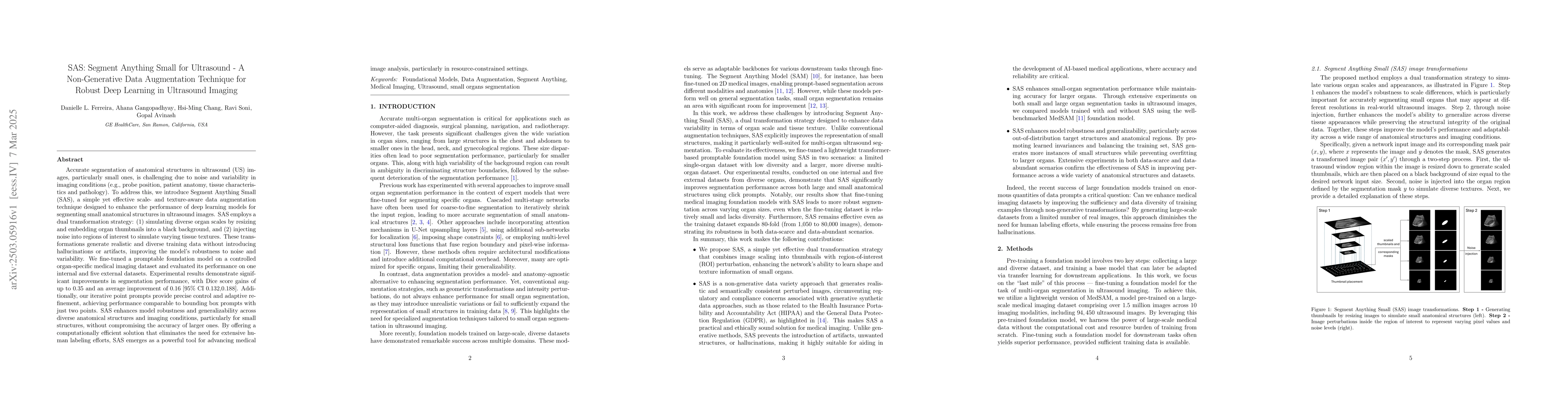
transformation strategy: (1) simulating diverse organ scales by resizing and embedding organ thumbnails into a black background, and (2) injecting noise into regions of interest to simulate varying tissue textures. These transformations generate realistic and diverse training data without introducing hallucinations or artifacts, improving the model's robustness to noise and variability. We fine-tuned a promptable foundation model on a controlled organ-specific medical imaging dataset and evaluated its performance on one internal and five external datasets. Experimental results demonstrate significant improvements in segmentation performance, with Dice score gains of up to 0.35 and an average improvement of 0.16 [95% CI 0.132,0.188]. Additionally, our iterative point prompts provide precise control and adaptive refinement, achieving performance comparable to bounding box prompts with just two points. SAS enhances model robustness and generalizability across diverse anatomical structures and imaging conditions, particularly for small structures, without compromising the accuracy of larger ones. By offering a computationally efficient solution that eliminates the need for extensive human labeling efforts, SAS emerges as a powerful tool for advancing medical image analysis, particularly in resource-constrained settings.
Key Findings, in focus
Seven facets of this paper, analysed and brought into focus by AI.
This research is important as it offers a computationally efficient solution to improve the segmentation of small anatomical structures in ultrasound images, which is crucial for medical diagnosis, especially in resource-constrained settings.
- SAS significantly improves segmentation performance with Dice score gains of up to 0.35 and an average improvement of 0.16 [95% CI 0.132,0.188].
- Iterative point prompts provide precise control and adaptive refinement, achieving performance comparable to bounding box prompts with just two points.
- SAS enhances model robustness and generalizability across diverse anatomical structures and imaging conditions, particularly for small structures, without compromising accuracy for larger ones.
This research is important as it offers a computationally efficient solution to improve the segmentation of small anatomical structures in ultrasound images, which is crucial for medical diagnosis, especially in resource-constrained settings.
The main technical contribution is the development of SAS, a novel, non-generative data augmentation technique that enhances the segmentation of small anatomical structures in ultrasound images by simulating scale variations and tissue textures.
SAS differs from existing methods by avoiding the introduction of artifacts, unwanted structures, or hallucinations, making it highly suitable for medical applications where accuracy and reliability are critical. It simulates expected domain shifts for specific anatomies by applying SAS to a single source domain, enabling deep models trained with SAS to better generalize to unseen domains.
- The study was conducted using ultrasound images and may not directly translate to other imaging modalities without further validation.
- The performance of SAS was evaluated on specific datasets, and its generalizability to a broader range of ultrasound images and anatomical structures requires further investigation.
- Applying SAS to other imaging modalities and integrating it into clinical workflows to validate its utility in real-world medical scenarios.
- Conducting a rigorous analysis of augmentation strength by varying parameters for image scaling, noise intensity, and the probability of applying perturbations during training.
Impact
Paper Details
Authors
PDF Preview
Citation Network
Current paper (gray), citations (green), references (blue)
Display is limited for performance on very large graphs.
Discussion 0