Authors
Summary
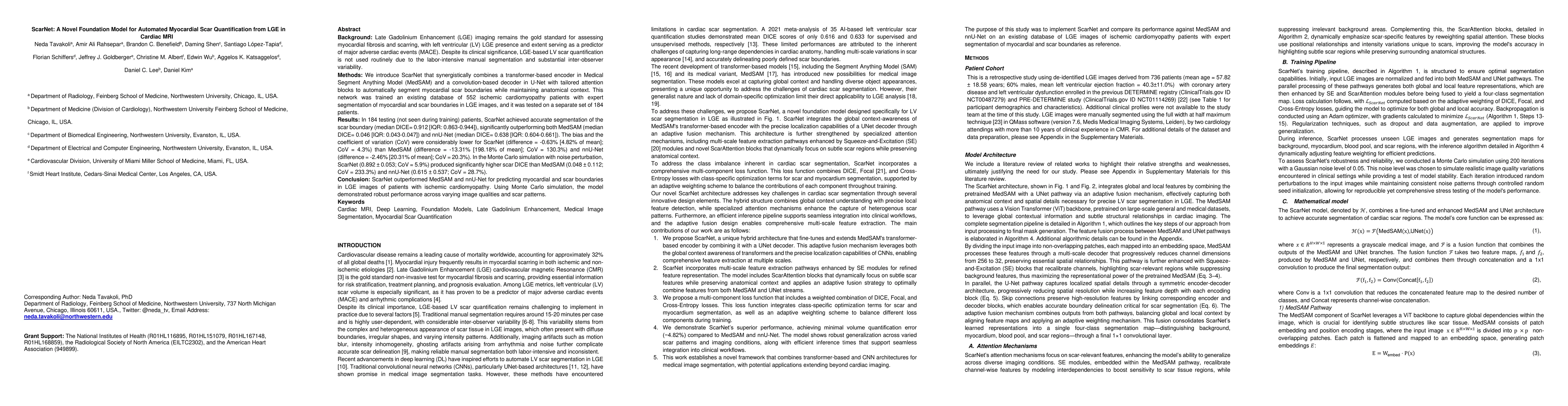
Background: Late Gadolinium Enhancement (LGE) imaging is the gold standard for assessing myocardial fibrosis and scarring, with left ventricular (LV) LGE extent predicting major adverse cardiac events (MACE). Despite its importance, routine LGE-based LV scar quantification is hindered by labor-intensive manual segmentation and inter-observer variability. Methods: We propose ScarNet, a hybrid model combining a transformer-based encoder from the Medical Segment Anything Model (MedSAM) with a convolution-based U-Net decoder, enhanced by tailored attention blocks. ScarNet was trained on 552 ischemic cardiomyopathy patients with expert segmentations of myocardial and scar boundaries and tested on 184 separate patients. Results: ScarNet achieved robust scar segmentation in 184 test patients, yielding a median Dice score of 0.912 (IQR: 0.863--0.944), significantly outperforming MedSAM (median Dice = 0.046, IQR: 0.043--0.047) and nnU-Net (median Dice = 0.638, IQR: 0.604--0.661). ScarNet demonstrated lower bias (-0.63%) and coefficient of variation (4.3%) compared to MedSAM (bias: -13.31%, CoV: 130.3%) and nnU-Net (bias: -2.46%, CoV: 20.3%). In Monte Carlo simulations with noise perturbations, ScarNet achieved significantly higher scar Dice (0.892 \pm 0.053, CoV = 5.9%) than MedSAM (0.048 \pm 0.112, CoV = 233.3%) and nnU-Net (0.615 \pm 0.537, CoV = 28.7%). Conclusion: ScarNet outperformed MedSAM and nnU-Net in accurately segmenting myocardial and scar boundaries in LGE images. The model exhibited robust performance across diverse image qualities and scar patterns.
AI Key Findings
Get AI-generated insights about this paper's methodology, results, and significance.
Paper Details
PDF Preview
Similar Papers
Found 4 papersJoint Deep Learning for Improved Myocardial Scar Detection from Cardiac MRI
Shuo Wang, Miaomiao Zhang, Jiarui Xing et al.
Robust Deep Learning for Myocardial Scar Segmentation in Cardiac MRI with Noisy Labels
Mostafa Mehdipour Ghazi, Gerry P. McCann, Evgeny M. Mirkes et al.
Contrast-Free Myocardial Scar Segmentation in Cine MRI using Motion and Texture Fusion
Lei Li, Guang Yang, Vicente Grau et al.
CLAIM: Clinically-Guided LGE Augmentation for Realistic and Diverse Myocardial Scar Synthesis and Segmentation
Chen Chen, Farheen Ramzan, Yusuf Kiberu et al.
No citations found for this paper.
Comments (0)